. 2009 Jan;20(1):114-22.
doi: 10.1681/ASN.2007111205. Epub 2008 Dec 10.

Adriamycin alters glomerular endothelium to induce proteinuria

Affiliations

Adriamycin alters glomerular endothelium to induce proteinuria

Marie Jeansson et al. J Am Soc Nephrol. 2009 Jan.

Abstract

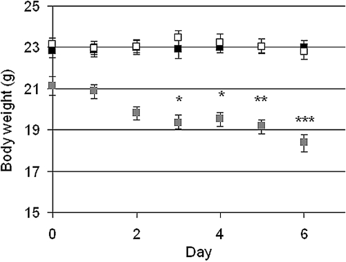
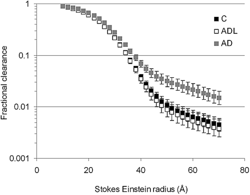
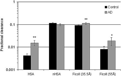
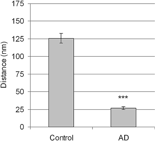
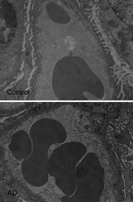
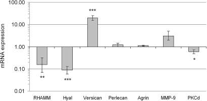
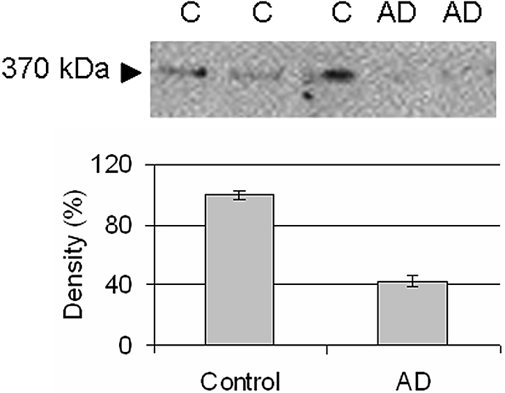
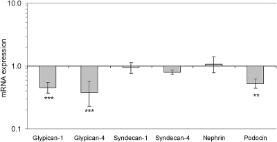
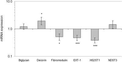
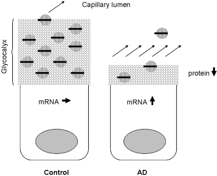
The pathophysiology underlying the nephrotic syndrome is becoming clear for several inherited podocytopathies; the mechanisms of injury that lead to the acquired forms of this disease are not well understood. We explored these mechanisms using the mouse model of adriamycin-induced proteinuria.We estimated the fractional clearances for FITC-Ficolls, albumin, and neutral albumin in cooled, isolated,perfused kidneys (cIPK) in situ. Treatment with adriamycin led to a significant increase in the fractional clearance of albumin and of Ficoll with radii larger than 20 A. Neutral albumin (33.4 A) and similarly sized Ficoll behaved similarly to each other. In addition, adriamycin led to a significant loss of charge density and size selectivity of the glomerular barrier. The thickness of the glomerular endothelial surface layer(i.e., or the glycocalyx) in adriamycin-treated animals was only 20% of that in normal animals. Finally,several proteoglycans were downregulated in isolated glomeruli. In summary, adriamycin thins the glomerular glycocalyx, perhaps by downregulating proteoglycan synthesis, and alters glomerular charge- and size selectivity. These data suggest that the glomerular endothelium may play a role in the pathogenesis of proteinuric renal diseases.


Figures

Figure 1.
Figure 1.
Mean ± SEM for the body weight for controls (black, n = 10), ADL (white, n = 9), and AD (gray, n = 9). Statistical analysis was made by comparing the body weights each day with the body weight at day 0. * P < 0.05, ** P < 0.01, *** P < 0.001.
Figure 2.
Figure 2.
Mean ± SEM for the fractional clearance of Ficolls (12 to 70Å) in controls and AD mice. There is a significant difference (P < 0.01) between controls (n = 10) and AD (n = 9) for all Ficolls. Note the logarithmic scale: only every 10th value is shown for clarity.
Figure 3.
Figure 3.
Mean ± SEM for the fractional clearance for HSA, nHSA, and Ficolls in controls (n = 10) and AD (n = 9) mice. Note the logarithmic scale. * P < 0.05, ** P < 0.01.
Figure 4.
Figure 4.
Mean ± SEM for the ESL in controls (four animals, n = 88 glomeruli) and AD mice (three animals, n = 79 glomeruli). *** P < 0.001.
Figure 5.
Figure 5.
Electron micrographs of glomerular capillaries showing Intralipid droplets and the podocyte foot-process flattening in AD mice.
Figure 6.
Figure 6.
Mean ± SEM for mRNA expression relative to housekeeping genes and controls (1) for RHAMM (hyaluronic acid motility receptor), hyaluronidase (Hyal), versican, perlecan, agrin, matrix metallopeptidase 9 (MMP-9), and protein kinase C δ (PKCδ) in AD mice. Note the logarithmic scale. n = 7 for controls and AD mice. * P < 0.05, ** P < 0.01 *** P < 0.001.
Figure 7.
Figure 7.
Western blot for versican (370 kD).
Figure 8.
Figure 8.
Mean ± SEM for mRNA expression relative to housekeeping genes and controls (1) for the heparan sulfate proteoglycans glypican-1, glypican-4, syndecan-1, syndecan-4, nephrin, and podocin. Note the logarithmic scale. n = 7 for controls and AD mice. *** P < 0.001.
Figure 9.
Figure 9.
Mean ± SEM for mRNA expression relative to housekeeping genes and controls (1) for the small leucine rich proteoglycans biglycan, decorin, and fibromodulin. Also shown are enzymes important for the elongation and sulfation of side chains (GAGs) on heparan sulfate proteoglycans EXT-1, HS2ST1, and NDST3. See Table 1 for abbreviations. Note the logarithmic scale. n = 7 for both controls and AD mice. * P < 0.05, ** P < 0.01, *** P < 0.001.
Figure 10.
Figure 10.
Speculative diagram of the versican mRNA and protein expression. In the normal endothelial cell, versican (in black) is produced and secreted into the ESL. Because of its size (approximately 1000 kD with GAG chains) and binding partners, versican has a slow turnover and a long diffusion distance to the plasma. However, in AD mice the glycocalyx thickness is decreased to approximately 20% of that in control mice and there is a loss of potential binding partners for versican. The consequence of this will be an increased loss of protein, for which the cell tries to compensate by increasing the translation of versican.

References

    1. Haraldsson B, Nyström J, Deen WM: Properties of the glomerular barrier and mechanisms of proteinuria. Physiol Rev 88: 451–487, 2008 - PubMed
    1. Comper WD, Haraldsson B, Deen WM: Resolved: Normal glomeruli filter nephrotic levels of albumin. J Am Soc Nephrol 19: 427–432, 2008 - PubMed
    1. Russo LM, Sandoval RM, McKee M, Osicka TM, Collins AB, Brown D, Molitoris BA, Comper WD: The normal kidney filters nephrotic levels of albumin retrieved by proximal tubule cells: Retrieval is disrupted in nephrotic states. Kidney Int, 71: 504–513, 2007 - PubMed
    1. Roselli S, Gribouval O, Boute N, Sich M, Benessy F, Attie T, Gubler MC, Antignac C: Podocin localizes in the kidney to the slit diaphragm area. Am J Pathol 160: 131–139, 2002 - PMC - PubMed
    1. Ruotsalainen V, Ljungberg P, Wartiovaara J, Lenkkeri U, Kestilä, M, Jalanko H, Holmberg C, Tryggvason, K: Nephrin is specifically located at the slit diaphragm of glomerular podocytes. Proc Natl Acad Sci U S A 96: 7962–7967, 1999 - PMC - PubMed

Publication types

LinkOut - more resources