. 2009 Feb;20(3):1089-101.
doi: 10.1091/mbc.e08-06-0566. Epub 2008 Dec 10.

Regulation of renal epithelial tight junctions by the von Hippel-Lindau tumor suppressor gene involves occludin and claudin 1 and is independent of E-cadherin

Affiliations

Regulation of renal epithelial tight junctions by the von Hippel-Lindau tumor suppressor gene involves occludin and claudin 1 and is independent of E-cadherin

Sarah K Harten et al. Mol Biol Cell. 2009 Feb.

Abstract

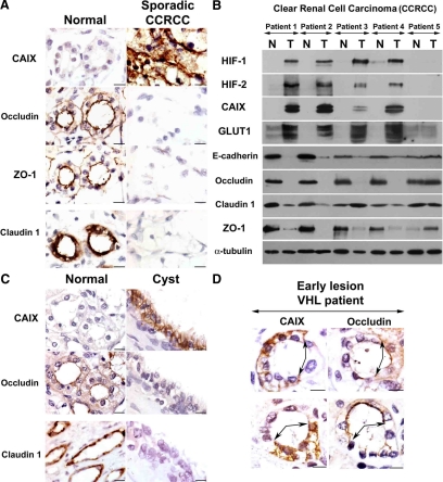
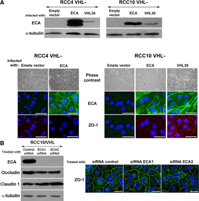
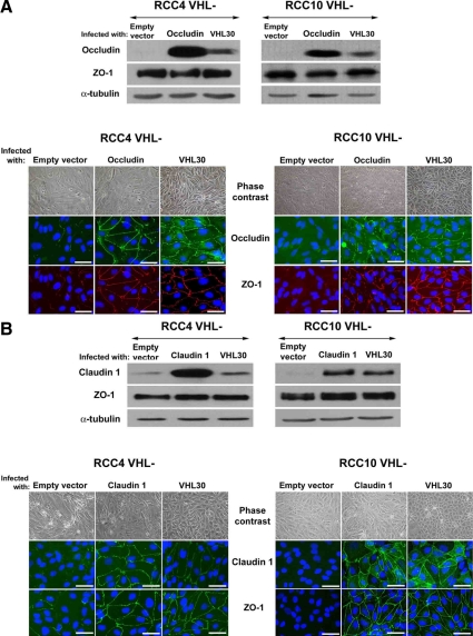
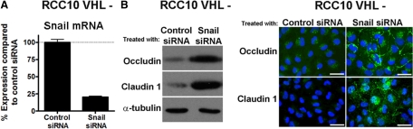
Epithelial-to-mesenchymal transitions (EMT) are important in renal development, fibrosis, and cancer. Loss of function of the tumor suppressor VHL leads to many features of EMT, and it has been hypothesized that the pivotal mediator is down-regulation of the adherens junction (AJ) protein E-cadherin. Here we show that VHL loss-of-function also has striking effects on the expression of the tight junction (TJ) components occludin and claudin 1 in vitro in VHL-defective clear cell renal cell carcinoma (CCRCC) cells and in vivo in VHL-defective sporadic CCRCCs (compared with normal kidney). Occludin is also down-regulated in premalignant foci in kidneys from patients with germline VHL mutations, consistent with a contribution to CCRCC initiation. Reexpression of E-cadherin was sufficient to restore AJ but not TJ assembly, indicating that the TJ defect is independent of E-cadherin down-regulation. Additional experiments show that activation of hypoxia inducible factor (HIF) contributes to both TJ and AJ abnormalities, thus the VHL/HIF pathway contributes to multiple aspects of the EMT phenotype that are not interdependent. Despite the independent nature of the defects, we show that treatment with the histone deacetylase inhibitor sodium butyrate, which suppresses HIF activation, provides a method for reversing EMT in the context of VHL inactivation.


Figures

Figure 1.
Figure 1.
VHL loss-of-function down-regulates the expression of occludin and claudin 1 in CCRCC cell lines. (A) Western blot showing that occludin and claudin 1 protein levels are increased when VHL is expressed in RCC4 and RCC10 cells. HIF-1α, HIF-2α, E-cadherin, ZO-1, and α-tubulin (loading control) are also shown. (B) Phase-contrast captures and immunofluorescence labeling for ZO-1, occludin, and claudin 1, in RCC4 and RCC10 cells. Labeling for claudin 1 in RCC4 cells was less intense than in RCC10. Bar, 40 μm. (C) Quantitative real-time RT-PCR analysis of occludin, claudin 1, ZO-1, and GLUT1 mRNA, in RCC4 and RCC10 cells and corresponding stable transfectants expressing VHL. Three independent experiments and the corresponding mean are presented. Differences in occludin, claudin 1, and GLUT1 mRNA expression levels were statistically significant (p < 0.05).
Figure 2.
Figure 2.
TJs are disrupted after VHL inactivation in renal-derived cells in vivo. (A) Serial sections from a representative sporadic VHL-defective CCRCC, which also includes adjacent unaffected tissue. Labeling was performed for ZO-1, occludin, claudin 1, and CAIX. Micrographs show representative fields of normal and tumor tissue from different areas of the same section. (B) Western blot with the indicated antibodies of five sporadic VHL-defective CCRCC tumors and paired unaffected kidney tissue. N, normal tissue; T, tumor. Consistent with VHL inactivation in the majority of sporadic CCRCCs, four of five tumors showed HIF activation (patients 1–4). E-cadherin, occludin, and ZO-1 were down-regulated in all four tumors displaying HIF activation; results with claudin 1 were more variable. (C) Serial sections labeled with the indicated antibodies of kidney tissue containing a cyst from a VHL patient (images are presented as in A). Ten independent cysts were analyzed with similar results. (D) Serial sections containing an early lesion of VHL inactivation (in an otherwise normal tubule) labeled for the HIF target gene CAIX and occludin. Early lesions from five VHL patients were analyzed and showed similar results. Bar, 10 μm.
Figure 3.
Figure 3.
VHL regulates TJ assembly independently of E-cadherin. (A) Western blot for E-cadherin using lysates from VHL negative RCC10 and RCC4 cells overexpressing E-cadherin; VHL 30 and empty vector were used as controls. Phase-contrast captures and immunofluorescence (double with ZO-1) of the same cell lines is also shown. E-cadherin was below the immunofluorescence detection threshold in the pool of RCC4 cells infected with VHL 30. (B) Western blot analysis of E-cadherin, occludin, and claudin 1 in RCC10/VHL cells treated with either a control oligo or two independent E-cadherin siRNA oligos. Representative phase-contrast photomicrographs and immunofluorescence captures of ZO-1 assembly are also shown. Bar, 40 μm. ECA, E-cadherin.
Figure 4.
Figure 4.
Reexpression of occludin or claudin 1 restores TJs in VHL negative CCRCC cells. VHL-defective RCC4 and RCC10 cell pools infected with retroviruses encoding (A) occludin, or (B) claudin 1. Empty vector and VHL 30 were used as controls. Immunofluorescence and phase contrast captures, and Western blots with the indicated antibodies are shown. Double labeling for ZO-1 and occludin is presented in A, and single labeling for claudin 1 and ZO-1 is shown in B. Bar, 40 μm.
Figure 5.
Figure 5.
Type IIC VHL mutations restore epithelial characteristics in VHL-defective RCC10 and RCC4 cells. Quantitative real-time RT-PCR analysis of PHD3 mRNA confirms suppression of HIF in VHL-defective RCC10 (A) and RCC4 (D) pools infected with VHL 19, VHL 30, and VHL V84L and VHL L188V. Three independent experiments and the corresponding mean are presented; differences were statistically significant (p < 0.05). (B) Phase-contrast images of RCC10 cell pools and immunofluorescence for ZO-1, occludin, and claudin 1. Bar, 40 μm. (C) Western blot for occludin, claudin 1, and α-tubulin in RCC10 cell pools. (E) Phase-contrast images of RCC4 cell pools and immunofluorescence for ZO-1 and occludin. Bar, 40 μm. (F) Western blot for occludin and α-tubulin in RCC4 cell pools.
Figure 6.
Figure 6.
HIF-1α is involved in the EMT-like phenotype of VHL-negative RCC10 cells. (A) Western blot with the indicated antibodies of VHL-defective RCC10 cells treated with control siRNA oligos, and siRNA oligos specific for HIF-1α or HIF-2α. A representative experiment is shown. Phase-contrast images and immunofluorescence labeling for ZO-1, occludin, and claudin 1 from the same siRNA-transfected cells. Immunofluorescence captures comparing different siRNA treatments were taken using similar exposure times. Quantification analyses of junction assembly was also performed across nine independent fields (∼50 cells per field) to permit comparison of the effects of silencing the individual HIF-α subunits. Error bars, SEM. (B) Western blot for HIF-1α and occludin, phase-contrast captures, and immunofluorescences for ZO-1 and occludin of VHL-positive RCC10 cells infected with a constitutively active form of HIF-1α. A representative experiment is shown. Bar, 40 μm.
Figure 7.
Figure 7.
SNAIL1 is involved in the EMT-like phenotype of VHL-negative RCC10 cells. (A) Quantitative real-time RT-PCR analysis of SNAIL1 mRNA levels in VHL-defective RCC10 cells treated with either control siRNA oligos or siRNA oligos specific for SNAIL1. Three independent experiments and the corresponding mean are presented. Snail1 knockdown was statistically significant (p < 0.05). (B) Representative Western blot and immunofluorescence captures show the expression level and TJ labeling of occludin and claudin 1 in RCC10 cell treated with SNAIL1 siRNA oligos. Bar, 40 μm.
Figure 8.
Figure 8.
Sodium butyrate improves epithelial characteristics in VHL-defective RCC4 and RCC10 cells. VHL-defective RCC4 (A) and RCC10 cells (B) were grown to confluence and treated for 3 d with the indicated doses of sodium butyrate, before phase-contrast imaging and immunofluorescence labeling, or immunoblotting. RCC4 and RCC10 cells stably expressing VHL were included for comparison in the immunoblot analysis. Bars, 40 μm.

Similar articles

Cited by

References

    1. Balkovetz D. F. Claudins at the gate: determinants of renal epithelial tight junction paracellular permeability. Am. J. Physiol. Renal Physiol. 2006;290:F572–F579. - PubMed
    1. Bi G., Jiang G. The molecular mechanism of HDAC inhibitors in anticancer effects. Cell Mol. Immunol. 2006;3:285–290. - PubMed
    1. Bilton R., Trottier E., Pouyssegur J., Brahimi-Horn M. C. ARDent about acetylation and deacetylation in hypoxia signalling. Trends Cell Biol. 2006;16:616–621. - PubMed
    1. Boutet A., De Frutos C. A., Maxwell P. H., Mayol M. J., Romero J., Nieto M. A. Snail activation disrupts tissue homeostasis and induces fibrosis in the adult kidney. EMBO J. 2006;25:5603–5613. - PMC - PubMed
    1. Braga V. M. Cell-cell adhesion and signalling. Curr. Opin. Cell Biol. 2002;14:546–556. - PubMed

Publication types

MeSH terms

LinkOut - more resources