. 2009 Feb;20(3):1006-19.
doi: 10.1091/mbc.e08-02-0140. Epub 2008 Dec 10.

Analysis of quality control substrates in distinct cellular compartments reveals a unique role for Rpn4p in tolerating misfolded membrane proteins

Affiliations

Analysis of quality control substrates in distinct cellular compartments reveals a unique role for Rpn4p in tolerating misfolded membrane proteins

Meredith Boyle Metzger et al. Mol Biol Cell. 2009 Feb.

Abstract

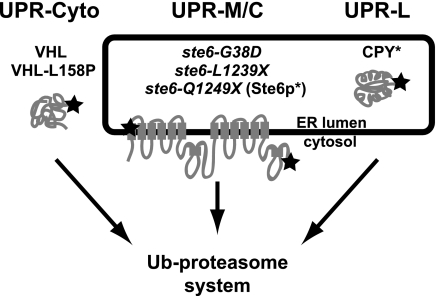
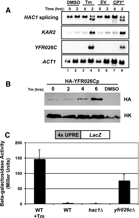
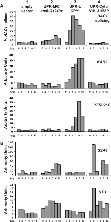
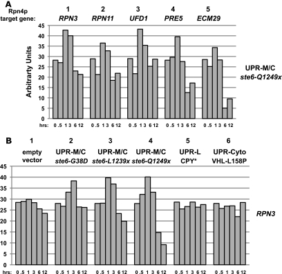
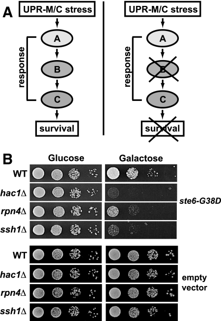
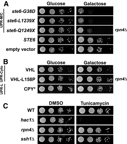
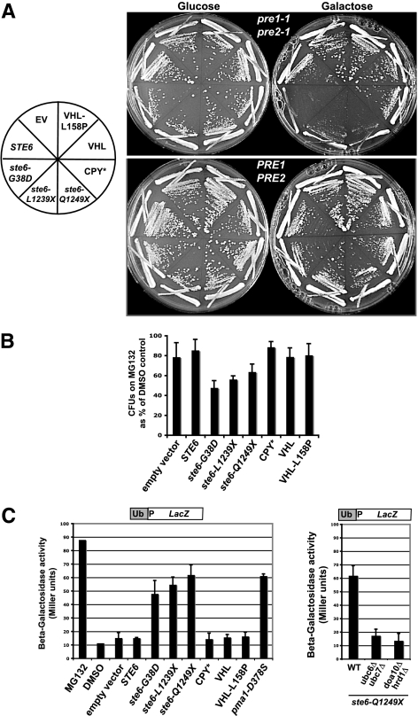
ER quality control (ERQC) prevents the exit of misfolded secretory and membrane proteins from the ER. A critical aspect of ERQC is a transcriptional response called the unfolded protein response (UPR), which up-regulates genes that enable cells to cope with misfolded, ER-retained proteins. In this study, we compare the transcriptional responses in yeast resulting from the acute expression of misfolded proteins residing in three different cellular compartments (the ER lumen, membrane, and cytosol), and find that each elicits a distinct transcriptional response. The classical UPR response, here-designated UPR-L, is induced by the ER lumenal misfolded protein, CPY*. The UPR-Cyto response is induced by the cytosolic protein, VHL-L158P, and is characterized by a rapid, transient induction of cytosolic chaperones similar to the heat-shock response. In contrast, the misfolded membrane protein with a cystolic lesion, Ste6p*, elicits a unique response designated UPR-M/C, characterized by the modest induction of >20 genes regulated by Rpn4p, an activator of proteasomal genes. Independently, we identified several genes required for yeast viability during UPR-M/C stress, but not UPR-L or UPR-Cyto stress. Among these is RPN4, highlighting the importance of the Rpn4p-dependent response in tolerating UPR-M/C stress. Further analysis suggests the requirement for Rpn4p reflects severe impairment of the proteasome by UPR-M/C stress.


Figures

Figure 1.
Figure 1.
Working model for three compartmentally distinct branches of the UPR. In this study, we ask if three classes of misfolded proteins with different cellular locations (ER lumen, membrane, and cytosol) induce distinct patterns of transcriptional responses. Misfolded proteins in the ER lumen, such as CPY*, induce the well-documented UPR-L transcriptional response (generally referred to simply as UPR); misfolded ER membrane proteins with membrane or cytosolic lesions, such as ste6-Q1249X (Ste6p*) and other Ste6p alleles (ste6-L1239X and ste6-G38D), could induce a UPR-M/C response; and misfolded proteins in the cytosol, such as VHL and VHL-L158P, could induce a UPR-Cyto response. All three classes of misfolded proteins are known to be degraded via the ubiquitin–proteasome system (Hiller et al., 1996; Loayza et al., 1998; McClellan et al., 2005). The star in the first transmembrane domain of Ste6p represents the site of the G38D mutation and the star in the C-terminal cytosolic domain represents the sites of the L1239X and Q1249X mutations.
Figure 2.
Figure 2.
YFR026C is a novel gene (designated ULI1, see discussion), highly induced by UPR-L stress. (A) Northern blot to examine UPR-L gene induction by the ER stressors, tunicamycin (Tm; lanes 1–4) and CPY* (lanes 5–8). In lanes 1–4, wild-type cells (SM4460) were treated with either DMSO or 10 μg/ml Tm. Samples were harvested before (0 h) and after (2 h) treatment. In lanes 5–8, wild-type cells (SM4460) expressing empty vector (EV; pSM922) or CPY* (pSM2215) were harvested before (0 h) and after (2 h) galactose induction. Blots were probed to examine HAC1 splicing, KAR2 induction, and YFR026C induction. The ACT1 levels served as a loading control. (B) A Western blot for YFR026Cp protein levels was performed using extracts prepared from cells expressing genomic HA-tagged YFR026Cp (SM5295) under control of its endogenous promoter. Cultures were either untreated (0 h) or treated with 10 μg/ml Tm for 2, 4, and 6 h or DMSO for 6 h and harvested for protein at each time point. Blots were probed with anti-HA and anti-Hexokinase (HK) antibodies. (C) To examine whether lack of YFR026C causes induction of the UPR-L, yfr026cΔ (SM5476) cells expressing a UPRE-LacZ reporter (pPW344) were assayed for β-galactosidase activity. For comparison, the activity of wild-type (SM4460) cells alone (WT) or treated with 10 μg/ml Tm for 2 h before processing (WT + Tm), and hac1Δ cells (SM5382), all expressing a UPRE-LacZ reporter (pPW344), are included as controls. The data reflect an average of three independent experiments; error bars, 1 SD.
Figure 3.
Figure 3.
Three distinct classes of misfolded proteins induce unique patterns of transcription. Cells expressing galactose-inducible constructs were harvesting for RNA processing and Northern blotting at 0, 0.5, 1, 3, 6, and 12 h after galactose induction. Shown is a representative experiment using the wild-type strain (SM4460) expressing empty vector (pSM922), ste6-Q1249X (pSM2213), CPY* (pSM2215), and VHL-L158P (pESC-L158P). (A) The indicated probes detect the UPR-L genes, KAR2, YFR026C, and HAC1, splicing. (B) Probes detect the cytosolic chaperones, SSA4 and STI1. Northern blots were performed in duplicate and shown is a representative experiment in which quantitated, normalized values, determined as described in Materials and Methods, are graphed as percent of HAC1 spliced or arbitrary units.
Figure 4.
Figure 4.
The UPR-M/C response is characterized by the induction of Rpn4p-target genes. (A) Wild-type cells (SM4460) expressing galactose-inducible ste6-Q1249X (pSM2213) were harvested for RNA processing at 0, 0.5, 1, 3, 6, and 12 h after galactose induction, and Northern blotting was performed with probes to the indicated UPR-M/C genes. (B) Northern blot analysis of wild-type cells (SM4460) expressing galactose-inducible empty vector (pSM922), ste6-G38D (pSM1898), ste6-L1239X (pSM2212), ste6-Q1249X (pSM2213), CPY* (pSM2215), or VHL-L158P (pESC-L158P) were harvested for RNA processing and Northern blotting with RPN3 at the indicated times after galactose induction. Northern blots were performed in duplicate and shown is a representative experiment in which quantitated, normalized values, determined as described in Materials and Methods, are graphed as arbitrary units.
Figure 5.
Figure 5.
Synthetic lethality analysis reveals a requirement for Rpn4p, Hac1p, and Ssh1p for viability during UPR-M/C stress. (A) Model for the use of synthetic lethality to identify genes in a pathway required to cope with a UPR-M/C stress. Hypothetical genes (labeled A–C and here are shown in a single pathway, for simplicity) are hypothesized to be required for survival of a UPR-M/C stress. When any of these genes are deleted, cells are inviable. (B) Serial dilutions of the wild-type (SM4460) and indicated mutant (hac1Δ, rpn4Δ, ssh1Δ; SM5382, SM5383, SM5384, respectively) strains containing the galactose-inducible ste6-G38D plasmid (pSM1898) or empty vector (pSM922) growing on the indicated media.
Figure 6.
Figure 6.
Rpn4p is required to survive UPR-M/C stress, but not UPR-L or UPR-Cyto stress. (A) Serial dilutions of the rpn4Δ mutant (SM5383) containing wild-type STE6 (pSM1897), empty vector (pSM922), or the UPR-M/C stresses ste6-G38D (pSM1898), ste6-L1239X (pSM2212), or ste6-Q1249X (pSM2213) are shown. (B) Serial dilutions of the rpn4Δ mutant (SM5383) containing plasmids that induce the UPR-Cyto stress, VHL (pESC-VHL) or VHL-L158P (pESC-L158P), or the UPR-L stress, CPY* (pSM2215) are shown. (C) Serial dilutions of wild-type (SM4460), rpn4Δ (SM5383), hac1Δ (SM5382), and ssh1Δ (SM5384) cells were grown on media containing 0.1 μg/ml tunicamycin or an equal volume DMSO for 2 d at 30°C.
Figure 7.
Figure 7.
Proteasome function is specifically impaired by the presence of misfolded membrane proteins. (A) Wild-type (PRE1 PRE2; WCG4a) and proteasome mutant strain pre1-1 pre2-1 (WCG4A-11/21A) containing the indicated galactose-inducible misfolded proteins (see Table 2 for plasmids names) were streaked to the indicated media and grown at 24°C for 4 d. (B) A strain sensitized to MG132, erg6::kanMX (SM4464), was transformed with plasmids expressing the indicated galactose-inducible misfolded proteins (see panel A). Transformants were assayed for viability by plating on solid media after an 8 h treatment in liquid with 50 μM MG132, as described in Materials and Methods. Colony-forming units (CFUs) were counted for MG132- and DMSO-treated cells and graphed as CFUs on MG132 as a percentage of CFUs on DMSO. Each experiment was repeated at least three times and graphed as the average; error bars, 1 SD. A paired t test gives p < 0.01 for ste6-G38D and a p < 0.05 for ste6-L1239X and ste6-Q1249X, all compared with empty vector. (C) To examine proteasome function, wild-type cells (SM4460) containing empty vector (pSM922) or the indicated galactose-inducible misfolded proteins and the Ub-Pro-LacZ reporter for proteasome activity (pSM2216) were induced by the addition of 4% galactose for 6 h at 30°C before preparation for β-galactosidase assay. For comparison, cells were treated with 50 μM MG132 or an equal volume of DMSO for 12 h at 30°C. Each experiment was repeated at least three times and graphed as the average; error bars, 1 SD. (D) To examine the affect of ubiquitination mutants on proteasome inhibition by ste6-Q1249X, wild-type (SM4460), ubc6Δ ubc7Δ (SM5362), and doa10Δ hrd1Δ (SM5360) cells expressing galactose-inducible ste6-Q1249X (pSM2213) and Ub-Pro-LacZ (pSM2216) were grown, prepared, and analyzed as in C.

References

    1. Apodaca J., Kim I., Rao H. Cellular tolerance of prion protein PrP in yeast involves proteolysis and the unfolded protein response. Biochem. Biophys. Res. Commun. 2006;347:319–326. - PubMed
    1. Bachmair A., Finley D., Varshavsky A. In vivo half-life of a protein is a function of its amino-terminal residue. Science. 1986;234:179–186. - PubMed
    1. Baker B. M., Tortorella D. Dislocation of an endoplasmic reticulum membrane glycoprotein involves the formation of partially dislocated ubiquitinated polypeptides. J. Biol. Chem. 2007;282:26845–26856. - PubMed
    1. Bence N. F., Sampat R. M., Kopito R. R. Impairment of the ubiquitin-proteasome system by protein aggregation. Science. 2001;292:1552–1555. - PubMed
    1. Bernales S., McDonald K. L., Walter P. Autophagy counterbalances endoplasmic reticulum expansion during the unfolded protein response. PLoS Biol. 2006;4:e423. - PMC - PubMed

Publication types

MeSH terms