. 2008 Dec 16;105(50):19762-7.
doi: 10.1073/pnas.0808393105. Epub 2008 Dec 10.

Beta-Catenin downregulation attenuates ischemic cardiac remodeling through enhanced resident precursor cell differentiation

Affiliations

Beta-Catenin downregulation attenuates ischemic cardiac remodeling through enhanced resident precursor cell differentiation

Laura C Zelarayán et al. Proc Natl Acad Sci U S A. .

Abstract

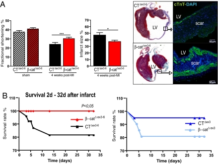
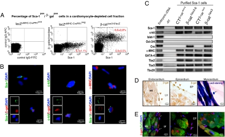
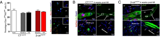
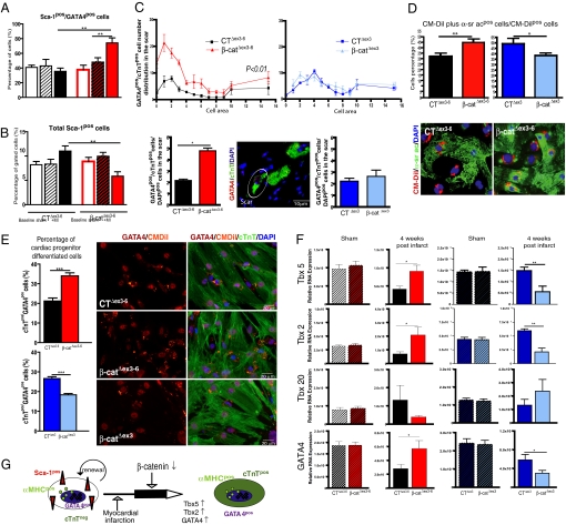
We analyzed the effect of conditional, alphaMHC-dependent genetic beta-catenin depletion and stabilization on cardiac remodeling following experimental infarct. beta-Catenin depletion significantly improved 4-week survival and left ventricular (LV) function (fractional shortening: CT(Deltaex3-6): 24 +/- 1.9%; beta-cat(Deltaex3-6): 30.2 +/- 1.6%, P < 0.001). beta-Catenin stabilization had opposite effects. No significant changes in adult cardiomyocyte survival or hypertrophy were observed in either transgenic line. Associated with the functional improvement, LV scar cellularity was altered: beta-catenin-depleted mice showed a marked subendocardial and subepicardial layer of small cTnT(pos) cardiomyocytes associated with increased expression of cardiac lineage markers Tbx5 and GATA4. Using a Cre-dependent lacZ reporter gene, we identified a noncardiomyocyte cell population affected by alphaMHC-driven gene recombination localized to these tissue compartments at baseline. These cells were found to be cardiac progenitor cells since they coexpressed markers of proliferation (Ki67) and the cardiomyocyte lineage (alphaMHC, GATA4, Tbx5) but not cardiac Troponin T (cTnT). The cell population overlaps in part with both the previously described c-kit(pos) and stem cell antigen-1 (Sca-1)(pos) precursor cell population but not with the Islet-1(pos) precursor cell pool. An in vitro coculture assay of highly enriched (>95%) Sca-1(pos) cardiac precursor cells from beta-catenin-depleted mice compared to cells isolated from control littermate demonstrated increased differentiation toward alpha-actin(pos) and cTnT(pos) cardiomyocytes after 10 days (CT(Deltaex3-6): 38.0 +/- 1.0% alpha-actin(pos); beta-cat(Deltaex3-6): 49.9 +/- 2.4% alpha-actin(pos), P < 0.001). We conclude that beta-catenin depletion attenuates postinfarct LV remodeling in part through increased differentiation of GATA4(pos)/Sca-1(pos) resident cardiac progenitor cells.


Conflict of interest statement

The authors declare no conflict of interest.

Figures

Fig. 1.
Fig. 1.
αMHC-dependent β-catenin depletion attenuates postinfarct LV remodeling. (A) β-Catenin depletion results in improved fractional shortening and reduced infarct size 4 weeks after infarct associated with a prominent subendocardial and subepicardial layer of cardiomyocytes as identified by cTnT staining. (B) Kaplan-Meier survival curve demonstrated significantly enhanced survival in β-catΔex3–6 mice compared to CTΔex3–6 mice 4 weeks following experimental infarct. In contrast, increased mortality was observed in β-catΔex3 mice compared to CTΔex3 mice. CTΔex3–6 n = 11; β-catΔex3–6 n = 15; CTΔex3 n = 8; β-catΔex3 n = 9; MI, myocardial infarct; LV, left ventricle; cTnT, cardiac Troponin T; *, P < 0.05.
Fig. 2.
Fig. 2.
Activation of the αMHC-promoter in cardiac progenitor cells. (A) Flow cytometry analysis reveals >80% of the noncardiomyocytes to coexpress β-gal and Sca-1 at baseline. Creneg mice were used as negative control. IgG-rabbit-APC and IgG-FITC antibodies were used as a control to discard unspecific staining. (B) Representative immunofluorescence picture of β-galpos cells from the cardiomyocyte depleted cell fraction proving coexpression of the Sca-1 and c-kit epitope. A subpopulation of cells coexpresses αMHC and Sca-1. (C) Analysis of Sca-1 cells gene expression by quantitative real-time PCR documenting expression of αMHC, c-kit, and cardiac transcription markers (GATA4, Tbx5). (D and E) Immunohistochemistry of adult heart from lacZαMHC-Cre mice. Activation of the αMHC promoter was analyzed by β-gal reporter gene expression. Small cells, with a high nuclei/cytoplasm ratio, positive for β-gal were detected by immunoperoxidase, enzymatic lacZ detection, and fluorescence staining in endo-, epi-, and myocardium.
Fig. 3.
Fig. 3.
Proliferation and tissue distribution of β-galpos/GATA4pos/cTnTneg cardiac progenitor cells after myocardial infarct. (A) Quantification of Sca-1pos/Ki67pos cell proliferation in a cardiomyocyte-depleted cell fraction by flow cytometry shows no significant differences between CTΔex3–6 and β-catΔex3–6 mice neither at baseline nor after ischemia. Immunofluorescence shows colocalization of Sca-1pos/BrdUpos and Sca-1pos/Ki67pos cells 2 weeks after infarct proving the proliferative capacity of these cells. (B and C) β-Catenin depleted β-catΔex3–6 mice exhibit a layer of cTnTpos cardiomyocytes along the scar, which appears less prominent in control mice (B and C, Upper Right). The subendocardial and subepicardial layer of the infarct scar is populated with small GATA4pos/cTnTneg cells (white arrows). In addition, small GATA4pos/cTnTpos cells are detected within the scar area of both β-catΔex3–6 and control mice (red arrows in B and C, Lower Left). No significant difference in tissue distribution is detected. LV, left ventricle; MI, myocardial infarct.
Fig. 4.
Fig. 4.
β-Catenin depletion enhances cardiomyocyte differentiation of Sca-1pos precursor cells following experimental infarct. (A) Significant increase of the Sca-1pos/GATA4pos cell fraction in β-catΔex3–6 mice 4 weeks after infarct vs. baseline and sham mice, which is not observed in CTΔex3–6 animals. It was accompanied by a nonsignificant decrease of total ScaIpos cells in β-catΔex3–6 mice while control mice tend to upregulate the total Sca-1 cells (B). (C) In Vivo quantification of GATA4pos/cTnTpos cells in the scar with a cell area <16 μm2 shows a significant increase of these cells in β-catΔex3–6 mice compared to controls. A representative confocal image is shown (white circle). (D) In Vitro increased differentiation of isolated Sca-1pos cells toward α-sr acpos cardiomyocytes from β-catΔex3–6 mice compared to isolated Sca-1 cells from CTΔex3–6 mice. The opposite effect was observed in β-catenin-stabilized mice. A representative picture of a 10-day CM-Dil-labeled coculture stained with α-sr ac is shown. (E) Similarly, in vitro differentiation of isolated Sca-1 cells toward GATA4pos/cTnTpos cells increases in β-catΔex3–6 compared to cells from CTΔex3–6 mice. The same cell population is decreased in β-catΔex3 mice. (F) Quantitative real-time PCR of heart samples 4 weeks after infarct shows an increased expression of markers for ventricular cardiomyocyte differentiation Tbx5, Tbx2, and GATA4 in β-catΔex3–6 mice compared to CTΔex3–6. Reciprocal regulation was observed in β-catΔex3 mice in comparison to CTΔex3 mice. (G) Hypothetical scheme of αMHCpos cardiac precursor cells differentiation in vivo triggered by ischemia and amplified by β-catenin depletion. *, P < 0.05; **, P < 0.001.

References

    1. Virag JA, et al. Fibroblast growth factor-2 regulates myocardial infarct repair. Effects on cell proliferation, scar contraction, and ventricular function. Am J Pathol. 2007;171:1431–1440. - PMC - PubMed
    1. Hsieh PCH, et al. Evidence from a genetic fate-mapping study that stem cells refresh adult mammalian cardiomyocytes after injury. Nat Med. 2007;13:970–974. - PMC - PubMed
    1. Oka T, Xu J, Molkentin JD. Re-employment of developmental transcription factors in adult heart disease. Sem Cell Dev Biol. 2007;18:117–131. - PMC - PubMed
    1. Oh H, et al. Cardiac progenitor cells from adult myocardium: Homing, differentiation, and fusion after infarction. Proc Natl Acad Sci USA. 2003;100:5834–5839. - PMC - PubMed
    1. Foley AC, Mercola M. Heart induction by Wnt antagonists depends on the homeodomain transcription factor Hex. Genes Dev. 2005;19:387–396. - PMC - PubMed

Publication types