. 2008 Dec 23;105(51):20404-9.
doi: 10.1073/pnas.0811011106. Epub 2008 Dec 10.

One-step assembly in yeast of 25 overlapping DNA fragments to form a complete synthetic Mycoplasma genitalium genome

Affiliations

One-step assembly in yeast of 25 overlapping DNA fragments to form a complete synthetic Mycoplasma genitalium genome

Daniel G Gibson et al. Proc Natl Acad Sci U S A. .

Abstract

We previously reported assembly and cloning of the synthetic Mycoplasma genitalium JCVI-1.0 genome in the yeast Saccharomyces cerevisiae by recombination of six overlapping DNA fragments to produce a 592-kb circle. Here we extend this approach by demonstrating assembly of the synthetic genome from 25 overlapping fragments in a single step. The use of yeast recombination greatly simplifies the assembly of large DNA molecules from both synthetic and natural fragments.


Conflict of interest statement

Conflict of interest statement: C.A.H., H.O.S., and J.C.V. have a financial interest in Synthetic Genomics, Inc (SGI). The research reported in this paper is in the field in which SGI is developing products. SGI has funded the research reported in the paper.

Figures

Fig. 1.
Fig. 1.
Construction of a synthetic M. genitalium genome in yeast. Yeast cells were transformed with 25 different overlapping A-series DNA segments (blue arrows; ≈17 kb to ≈35 kb each) composing the M. genitalium genome. To assemble these into a complete genome, a single yeast cell (tan) must take up at least one representative of the 25 different DNA fragments and incorporate them in the nucleus (yellow), where homologous recombination occurs. This assembled genome, called JCVI-1.1, is 590,011 bp, including the vector sequence (red triangle) shown internal to A86–89. The yeast propagation elements contained within the vector are an origin of replication (ARSH4), a centromere (CEN6), and a histidine-selectable marker (HIS3). In addition to full assembly of the genome as depicted here, some yeast cells may take up fewer than 25 different pieces and produce subassemblies of the genome by a mechanism such as NHEJ (see text). Others may take up more than 25 fragments and produce more than one assembled molecule per cell (not illustrated).
Fig. 2.
Fig. 2.
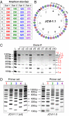
Multiplex PCR analysis to screen for yeast cells that took up all 25 segments. (A) Forty amplicons were designed such that 10 products could be produced in four separate multiplex PCR reactions (set 1 [red], set 2 [green], set 3 [blue], set 4 [purple]). PCR products ranged in size from 100 bp to 1075 bp and could be easily separated by electrophoresis on 2% agarose gels (C and D). (B) The 40 sets of primers amplified a small portion of the M. genitalium genome approximately once every 15 kb. Each of the 25 fragments (gray arrows) provided primer-binding sites for at least 1 of the 40 amplicons (red, green, blue, and purple lines). The vector sequence is represented by a black triangle. (C) DNA was extracted from 10 yeast clones, and multiplex PCR, with primer set 1, was performed. Clones 1 and 4 (c1 and c4) efficiently generated 9 of the 10 predicted amplicons. Clone 4 was selected for further analysis. (D) Multiplex PCR was performed on clone 4 and JCVI-1.0 as a positive control using all four primer sets. With the exception of amplicon 1-h, all 40 amplicons were efficiently generated from these clones. In the lanes labeled “L,” the 100-bp ladder (New England Biolabs) was loaded; sizes are indicated.
Fig. 3.
Fig. 3.
Validation of an intact M. genitalium genome by restriction analysis. (A) Diagram of the EagI (red), BssHII (blue), and AatII (green) restriction fragments expected for a complete and proper assembly of the synthetic JCVI-1.1 M. genitalium genome. The 25 pieces are represented by gray arrows. (B) The sizes of the five restriction fragments indicated in (A). (C) Total DNA from clone 4 (Y + Mg) and its host strain alone (Y) were isolated from yeast cells embedded in agarose. Most of the linear DNA was electrophoresed out of the agarose plugs. These plugs were then digested with EagI, BssHII, or AatII and analyzed by FIGE on 1% agarose gels. Restriction fragments corresponding to the correct sizes are indicated by the fragment numbers shown in (A) and (B). In the lanes labeled “λ,” the lambda ladder (New England Biolabs) was loaded; sizes are indicated.
Fig. 4.
Fig. 4.
PCR analysis of linearized and gel-purified JCVI-1.1. (A) Total DNA from JCVI-1.1 (c4), its host strain alone as a negative control, and JCVI-1.0 as a positive control were prepared and then analyzed after digestion with EagI as shown in Fig. 3. JCVI-1.0 has two additional EagI sites within its vector sequence, producing two genome fragments of ≈233 kb and ≈350 kb as well as a released ≈9-kb vector. The linearized JCVI-1.0 and JCVI-1.1 genome fragments were cut out of the gel, as indicated by red rectangles. (B) DNA from the gel slices in (A) was extracted and purified, then used as template for multiplex PCR, as shown in Fig. 2. (C) Twenty-five amplicons (J1-J25, black lines) are produced from 25 sets of primers that span each of the 25 junctions at which joining of the A-series assemblies (gray arrows) occurs. The predicted amplicon sizes for correctly assembled molecules are indicated. It was necessary to design larger amplicons for J08, J10, and J16 to ensure unique primer binding sites due to MgPa repeats at these junctions. (D) Purified JCVI-1.0 and JCVI-1.1 DNA extracted from the gel slices in (A) produced all 25 expected fragments after PCR. Products were analyzed on 2% agarose gels except for J08, J10, and J16, which were analyzed on 0.8% agarose gels. In the lanes labeled “M,” the 1-kb ladder (New England Biolabs) was loaded; sizes are indicated.
Fig. 5.
Fig. 5.
The construction of JCVI-1.9 by an independent 25-piece assembly. (A) DNA was extracted from 10 yeast clones (c11–c20), and multiplex PCR, with primer set 3, was performed. Clone 11 generated all 10 predicted amplicons and thus was selected for further analysis. (B) Multiplex PCR was performed on clone 11 using all four primer sets. All 40 amplicons were efficiently generated from this clone. (C) DNA was prepared from clone 11 in agarose plugs, then digested with EagI, as shown in Fig. 3. The linearized genome (≈587 kb) was separated on a 1% agarose gel in 0.5X Tris-borate-EDTA buffer at 14 °C on a Bio-Rad Mapper XA CHEF system. The run time was 16 h at 6 V/cm with a 50–90 s switch time ramp at an included angle of 120°. The linearized JCVI-1.9 genome fragment was cut out of the gel as indicated by the red rectangle. (D) DNA from the gel slice in (C) was extracted and purified, then used as template for PCR at the 25 junctions shown in Fig. 4. All 25 predicted amplicons were observed after analysis on 2% and 0.8% agarose gels.
Fig. 6.
Fig. 6.
Plot of the probability that an entire set of 25 DNA pieces is taken up (success) versus the number of DNA pieces taken up by an individual yeast cell. Data are from a computer simulation. N draws were made from a bag of 25 objects (pieces of DNA), replacing the object after each draw. The trial was a success if all 25 objects were drawn at least once. A million trials were conducted for each N to obtain a good approximation to the true probability of success.

References

    1. Hinnen A, Hicks JB, Fink GR. Transformation of yeast. Proc Natl Acad Sci U S A. 1978;75:1929–1933. - PMC - PubMed
    1. Orr-Weaver TL, Szostak JW, Rothstein RJ. Yeast transformation: A model system for the study of recombination. Proc Natl Acad Sci U S A. 1981;78:6354–6358. - PMC - PubMed
    1. Ma H, Kunes S, Schatz PJ, Botstein D. Plasmid construction by homologous recombination in yeast. Gene. 1987;58:201–216. - PubMed
    1. Raymond CK, Pownder TA, Sexson SL. General method for plasmid construction using homologous recombination. Biotechniques. 1999;26:134–141. - PubMed
    1. Raymond CK, Sims EH, Olson MV. Linker-mediated recombinational subcloning of large DNA fragments using yeast. Genome Res. 2002;12:190–197. - PMC - PubMed

Publication types

MeSH terms

LinkOut - more resources