. 2008 Dec 10;28(50):13368-83.
doi: 10.1523/JNEUROSCI.2918-08.2008.

Brain-derived neurotrophic factor signaling does not stimulate subventricular zone neurogenesis in adult mice and rats

Affiliations

Brain-derived neurotrophic factor signaling does not stimulate subventricular zone neurogenesis in adult mice and rats

Rui P Galvão et al. J Neurosci. .

Abstract

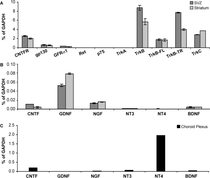
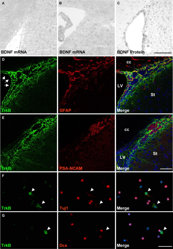
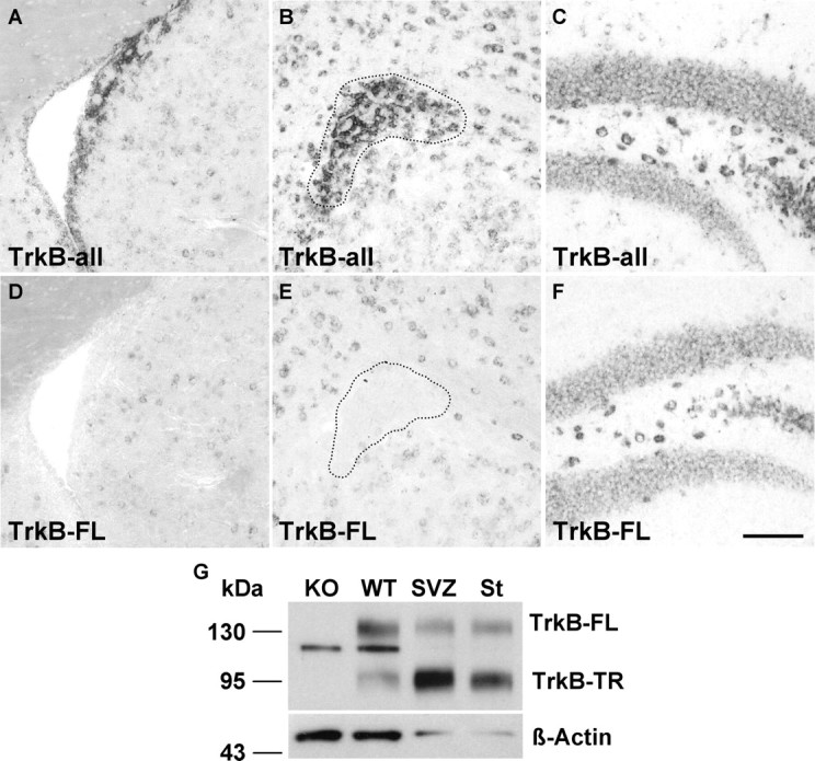
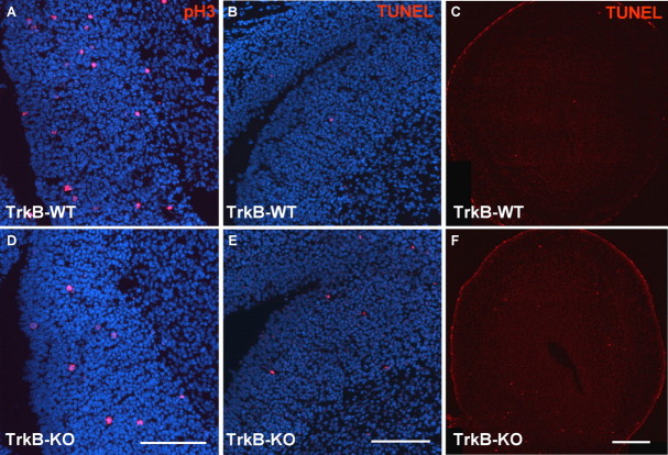
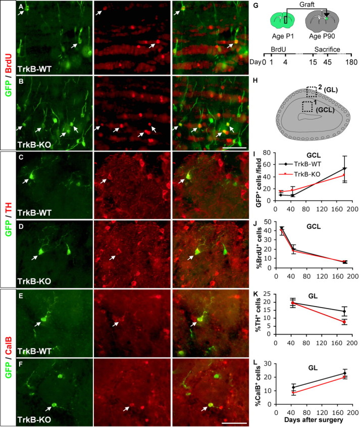
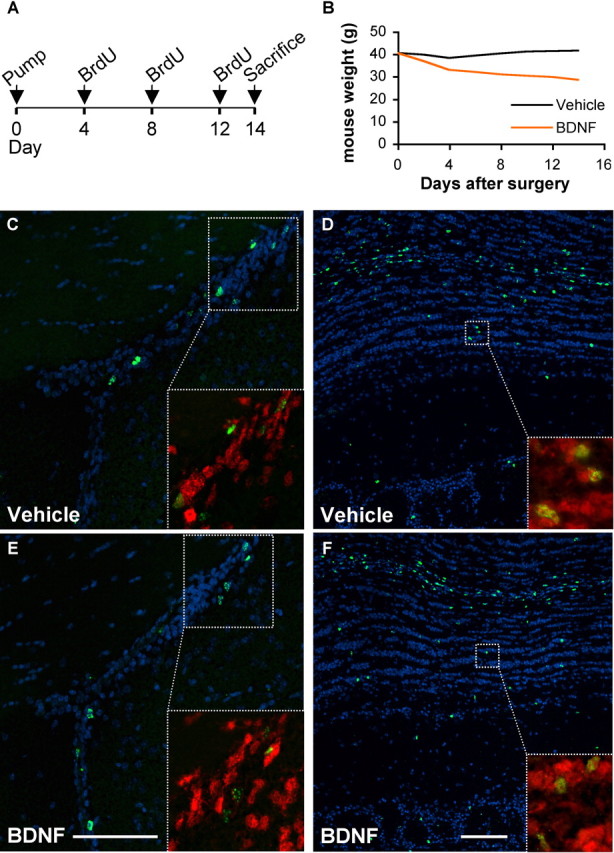
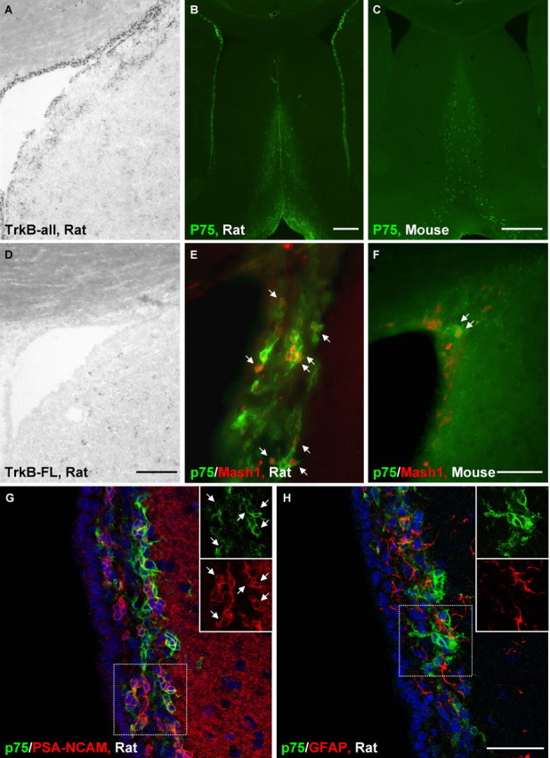
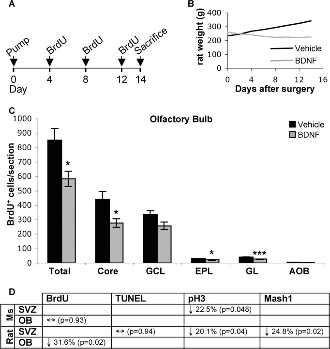
In rodents, the adult subventricular zone (SVZ) generates neuroblasts which migrate to the olfactory bulb (OB) and differentiate into interneurons. Recent work suggests that the neurotrophin Brain-Derived Neurotrophic Factor (BDNF) can enhance adult SVZ neurogenesis, but the mechanism by which it acts is unknown. Here, we analyzed the role of BDNF and its receptor TrkB in adult SVZ neurogenesis. We found that TrkB is the most prominent neurotrophin receptor in the mouse SVZ, but only the truncated, kinase-negative isoform (TrkB-TR) was detected. TrkB-TR is expressed in SVZ astrocytes and ependymal cells, but not in neuroblasts. TrkB mutants have reduced SVZ proliferation and survival and fewer new OB neurons. To test whether this effect is cell-autonomous, we grafted SVZ cells from TrkB knock-out mice (TrkB-KO) into the SVZ of wild-type mice (WT). Grafted progenitors generated neuroblasts that migrated to the OB in the absence of TrkB. The survival and differentiation of granular interneurons and Calbindin(+) periglomerular interneurons seemed unaffected by the loss of TrkB, whereas dopaminergic periglomerular neurons were reduced. Intra-ventricular infusion of BDNF yielded different results depending on the animal species, having no effect on neuron production from mouse SVZ, while decreasing it in rats. Interestingly, mice and rats also differ in their expression of the neurotrophin receptor p75. Our results indicate that TrkB is not essential for adult SVZ neurogenesis and do not support the current view that delivering BDNF to the SVZ can enhance adult neurogenesis.


Figures

Figure 1.
Figure 1.
RT-PCR analysis of neurotrophin receptors and ligands. We used semiquantitative RT-PCR to determine the relative expression levels of multiple neurotrophins and their receptors in the adult mouse brain (CNTFR, gp130: receptors for CNTF; GFRα1, Ret: receptors for GDNF; p75: low affinity receptor for NGF, NT3, NT4, BDNF; TrkA: high affinity receptor for NGF; TrkB: high affinity receptor for NT4, BDNF; TrkB-FL and TrkB-TR: full-length and truncated isoforms of TrkB receptor; TrkC: high affinity receptor for NT3). A, Neurotrophin receptor expression levels in SVZ and striatum. B, Neurotrophin expression levels in SVZ and striatum. C, neurotrophin expression levels in choroid plexus. All values are normalized to GAPDH expression and represent average ± SEM. Data in A, B, represent average from two experiments, pooling RNA from 8 adult mice/experiment; data in C, represents RNA of 16 adult mice pooled in one experiment.
Figure 2.
Figure 2.
Expression analysis of BDNF and TrkB in the mouse SVZ. A, B, In situ hybridization detects very low levels of BDNF mRNA at all levels of the adult SVZ (A, anterior SVZ; B, medial/posterior SVZ). C, Immunostaining detection of BDNF protein in the adult SVZ. D, E, Confocal images of TrkB immunostaining in adult mouse SVZ. D, TrkB (green) colocalizes with the astrocyte marker GFAP (red); arrows: ependymal cells lining the ventricular wall also express TrkB. E, TrkB (green) does not colocalize with the neuroblast marker PSA-NCAM (red). F, G, Immunostaining for TrkB receptor (green) on acutely dissociated neonatal SVZ cells confirms that SVZ neuroblasts, detected with Tuj1 (F, red) or Dcx antibodies (G, red) do not express TrkB (arrowheads: TrkB+ cells isolated from SVZ are negative for Tuj1 and Dcx). Scale bars: (A–C), 100 μm; (D, E), 50 μm; (F, G), 30 μm. cc, corpus callosum; LV, lateral ventricle; St, striatum.
Figure 3.
Figure 3.
Expression of TrkB isoforms in the adult mouse SVZ. A–C, In situ hybridization to detect all isoforms of TrkB (TrkB-all) shows strong staining in the SVZ (A), RMS (B) and hippocampal dentate gyrus (C). D–F, In situ hybridization to specifically detect the full-length isoform of TrkB (TrkB-FL) shows that SVZ (D) and RMS (E) cells do not express this isoform, although hippocampal cells (F) do. G, Western blot for TrkB shows that the predominant isoform in the SVZ is TrkB-TR at the protein level as well. Striatum protein was run for comparison and samples from TrkB-KO and WT whole brain (collected at P1) were used as controls; all samples were probed for β-Actin as loading control. Scale bar, 100 μm. Dashed line, outline of RMS.
Figure 4.
Figure 4.
In vivo consequences of TrkB-KO. P0 mice were stained in the SVZ and OB for phosphorylated histone H3 (pH3) and TUNEL to detect proliferating and apoptotic cells, respectively. A, D, pH3 immunostaining (red) in SVZ of TrkB-WT (A) and TrkB-KO (D) mice. B, E, TUNEL (red) in SVZ of TrkB-WT (B) and TrkB-KO (E) mice. C, F, TUNEL (red) in OB of TrkB-WT (C) and TrkB-KO (F) mice. Blue: DAPI (all panels). Scale bars: (A, D; B, E), 100 μm; (C, F), 1 mm.
Figure 5.
Figure 5.
Grafted TrkB-KO cells can survive and mature in OB. β-actin:GFP+ cells from SVZ of TrkB-WT or TrkB-KO P1 mice were grafted into the SVZ of WT, P90 mice; GFP+ cells were then analyzed in the OB 15, 45 and 180 d after surgery; BrdU was administered during the first 3 d after surgery. A, B, GFP/BrdU (green/red) staining in the granular cell layer (GCL) of the OB of mice grafted with TrkB-WT (A) or TrkB-KO (B) cells. C, D, GFP/TH (green/red) staining in the glomerular layer (GL) of the OB of mice grafted with TrkB-WT (C) or TrkB-KO (D) cells. E, F, GFP/CalB (green/red) staining in the GL of mice grafted with TrkB-WT (E) or TrkB-KO (F) cells. G, Experimental design. H, Diagram of a coronal section of OB (dorsal to the left), showing general location from which data were collected (box 1: origin of data in A, B, I, J; box 2: origin of data in C-F, K, L). I, Quantification of all GFP+ cells/field (field = 435 × 330 μm) in GCL of OB, showing increase in both TrkB-WT and TrkB-KO cells throughout entire experiment; n = 3 (15 d graft), n = 2 (45 d graft), n = 2 (180 d graft). J, Quantification of the proportion of grafted cells labeled with BrdU in GCL of OB, showing identical dynamics for TrkB-WT and TrkB-KO cells; n = 3 (15 d graft), n = 4 (45 d graft), n = 2 (180 d graft). K, Proportion of grafted cells expressing TH protein in GL of OB: there is a sharper drop in TH+ cells among TrkB-KO cells than among TrkB-WT cells between 45 and 180 d after surgery; n = 4 (all time points except 45 d TrkB-WT, for which n = 3, and 180 d TrkB-KO, for which n = 5). L, Proportion of grafted cells expressing CalB protein in GL of OB: both TrkB-WT and TrkB-KO grafted cells increase between 45 and 180 d after surgery; n = 4 (all time points except 45 d TrkB-KO, for which n = 3). All values represent average ± SEM. All images shown were taken at 45-day graft survival. Arrows indicate double-labeled cells. TH: tyrosine hydroxylase; CalB: Calbindin. Scale bars: (A, B), 100 μm; (C–F), 50 μm.
Figure 6.
Figure 6.
In vivo effect of BDNF infusion in adult mice. A, Experimental design: micro-osmotic pumps were implanted on P90 mice to infuse BDNF for 14 d; BrdU was administered every 4 d by intraperitoneal injection; on day 14, mice were killed for analysis. B, Effect of BDNF infusion on mouse weight. C, E, Phosphorylated histone H3 (pH3, green) staining ipsilateral to pump implant, in SVZ of vehicle (C) and BDNF (E) infused mice. D, F, BrdU (green) staining of OB ipsilateral to pump implant in vehicle (D) and BDNF (F) infused mice (see supplemental Fig. 12A, available at www.jneurosci.org as supplemental material, for BrdU quantification in all OB layers). SVZ inset (C, E), pH3/Hu (green/red); OB inset (D, F): BrdU/Hu (green/red). Blue: DAPI (all panels). Scale bars: 100 μm (C–F).
Figure 7.
Figure 7.
p75 but not TrkB receptor expression is different in adult rat and mouse SVZ. A, D, In situ hybridization to detect all isoforms of TrkB (TrkB-all, A) shows strong staining in the ependymal layer and in some cells of the adult rat SVZ, whereas the full-length isoform of TrkB (TrkB-FL, D), although present in striatal cells, is undetectable in the rat SVZ. B, p75 was detected by immunohistochemistry in many cells at all levels of the adult rat SVZ as well as in the medial septum. C, In contrast, very little p75 staining was detected in the adult mouse SVZ, although the medial septum contained again many p75+ cells. E, F, p75+ cells often coexpressed the transcription factor Mash1 (red) in both rat (E) and mouse (F) SVZ. G, H, Confocal microscopy (single image) of adult rat SVZ reveals significant coexpression of p75 (green) with the neuroblast marker PSA-NCAM (G, red) but not with the astrocyte marker GFAP (H, red). Insets show individual markers from boxed areas; arrows indicate several double-labeled cells (blue: DAPI). Scale bars: (A, D; E, F), 100 μm; (B, C), 500 μm; (G, H), 50 μm.
Figure 8.
Figure 8.
In vivo effect of BDNF infusion in adult rats. A, Experimental design: micro-osmotic pumps were implanted on ∼250 g rats to infuse BDNF or vehicle for 14 d; BrdU was administered every 4 d by intraperitoneal injection; on day 14, rats were killed for analysis. B, Effect of BDNF infusion on rat weight. C, Quantification of BrdU staining in rat OB ipsilateral to pump; results are presented for all OB layers together (Total) as well as for each individual layer (Core; GCL, granular cell layer; EPL, external plexiform layer; GL, glomerular layer; AOB, accessory olfactory bulb). D, Summary of quantifications for mouse (Ms) and rat BDNF infusion experiments (in both of which new OB cells were labeled with 3 BrdU injections, 4 d apart from each other); p values represent statistical significance by Student's t test; *p < 0.05, ***p < 0.001. n = 5 animals/group for all mouse quantifications; n = 6 animals/group for all rat quantifications, except BrdU (n = 5 Vehicle rats, 6 BDNF rats) and TUNEL (n = 6 Vehicle rats, 5 BDNF rats) quantifications. All values represent average ± SEM.

Similar articles

Cited by

References

    1. Ahmed S, Reynolds BA, Weiss S. BDNF enhances the differentiation but not the survival of CNS stem cell-derived neuronal precursors. J Neurosci. 1995;15:5765–5778. - PMC - PubMed
    1. Alcantara S, Frisen J, del Rio JA, Soriano E, Barbacid M, Silos-Santiago I. TrkB signaling is required for postnatal survival of CNS neurons and protects hippocampal and motor neurons from axotomy-induced cell death. J Neurosci. 1997;17:3623–3633. - PMC - PubMed
    1. Alvarez-Buylla A, Garcia-Verdugo JM. Neurogenesis in adult subventricular zone. J Neurosci. 2002;22:629–634. - PMC - PubMed
    1. Anderson KD, Alderson RF, Altar CA, DiStefano PS, Corcoran TL, Lindsay RM, Wiegand SJ. Differential distribution of exogenous BDNF, NGF, and NT-3 in the brain corresponds to the relative abundance and distribution of high-affinity and low-affinity neurotrophin receptors. J Comp Neurol. 1995;357:296–317. - PubMed
    1. Baker H, Kawano T, Margolis FL, Joh TH. Transneuronal regulation of tyrosine hydroxylase expression in olfactory bulb of mouse and rat. J Neurosci. 1983;3:69–78. - PMC - PubMed

Publication types

MeSH terms

Substances