. 2008 Dec 10;28(50):13411-8.
doi: 10.1523/JNEUROSCI.4095-08.2008.

Normal electrocortical facilitation but abnormal target identification during visual sustained attention in schizophrenia

Affiliations

Normal electrocortical facilitation but abnormal target identification during visual sustained attention in schizophrenia

Brett A Clementz et al. J Neurosci. .

Abstract

Attentional deficits in schizophrenia have been investigated using target identification tasks which conflate the abilities to successfully (1) attend to possible target locations and (2) detect target events. Whether compromised attentional selectivity or abnormal target detection causes schizophrenia subjects' poor performance on visual attention tasks, therefore, is unknown. To address this issue, we measured the neural activity (using electroencephalography) of 17 schizophrenia and 17 healthy subjects during a target identification task. Participants viewed superimposed images (horizontal and vertical bars differing in color) and attended to one image to identify bar width changes in specific locations. Bars were frequency tagged so attention directed to unique parts of the images could be tracked. Steady-state visual evoked potentials (ssVEPs) were used to quantify attention-related neural activity to specific parts of the visual images. Behavioral performance and event-related potentials (ERPs) in response to the target events were used to quantify target detection abilities. For both schizophrenia and healthy subjects, attending to specific parts of the attended image enhanced brain activity related to attended bars and reduced activity evoked by unattended bars. Activity in relation to the spatially overlapping unattended image was unaffected. Schizophrenia patients, however, were impaired on target detection abilities on both behavioral and brain activity measures. Target-related behavioral and brain activity measures were highly correlated in both groups. These findings indicate that deficient target detection rather than compromised attentional selectivity accounts for previously reported visual attention deficits in schizophrenia.


Figures

Figure 1.
Figure 1.
The two images (horizontal and vertical bars) used in this study are shown as spatially distinct, but they perfectly overlapped during testing. Each image included the middle bar, the peripheral bars, and the outside bars.
Figure 2.
Figure 2.
The top row of the figure shows the head surface map of grand average FFT power to the steady-state stimuli (averaged over all frequencies and all conditions for all participants) for both healthy (left) and schizophrenia (right) subjects. The bottom row of the figure shows the sLORETA solutions of time-domain averaged ssVEPs for both healthy and schizophrenia subjects.
Figure 3.
Figure 3.
An example of power spectra averaged over the 67 EEG channels that captured the peak ssVEP power (healthy subjects: black line; schizophrenia subjects: red line). In this example, the red vertical middle bar, peripheral bars, and outside bars flickered at 7.69 Hz, 7.14 Hz, and 6.67 Hz, respectively. The green horizontal bars flickered at 8.33 Hz. Participants attended to the red vertical bars, and were asked to identify width changes in the red middle and peripheral bars (A, the attend-all condition), red middle bar only (B, the attend-mid condition) or red peripheral bars only (C, the attend-peripheral condition).
Figure 4.
Figure 4.
Mean amplitude (plus SE) of the ssVEP for healthy and schizophrenia subjects in the different attention conditions. The labels on the x-axis refer to the different stimulation locations that had unique stimulation frequencies. Note that ssVEP amplitude to the unattended image is higher because there are more stimuli (5 total bars, so greater overall neural activity) contributing to this response.
Figure 5.
Figure 5.
The top portion of the figure shows the grand average VEP waveforms for healthy (black line) and schizophrenia (red line) subjects. They show separate averages for the VEPs to the attended objects (solid line), unattended objects (dashed line), and unattended image (light solid line), respectively. The back views of the head on the far left (healthy subjects) and right (schizophrenia patients) show the grand average voltage and surface laplacian topographies for the P3 component (same scale for both groups). The bottom middle image shows the sLORETA solution of the P3 amplitude difference between health and schizophrenia subjects, and indicates stronger activity among healthy persons specifically in the left superior parietal region.
Figure 6.
Figure 6.
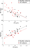
Bivariate scatter plot of relationships between behavioral responses (upper plot: reaction time; lower plot: d′) and P3 amplitude for healthy (black dots) and schizophrenia (red dots) subjects. The best fitting regression lines are also included for both groups.

Similar articles

Cited by

References

    1. American Psychiatric Association. Ed 4. Washington, DC: American Psychiatric Association; 1994. Diagnostic and statistical manual of mental disorders.
    1. Brody SA, Conquet F, Geyer MA. Disruption of prepulse inhibition in mice lacking mGluR1. Eur J Neurosci. 2003;18:3361–3366. - PubMed
    1. Butler PD, Martinez A, Foxe JJ, Kim D, Zemon V, Silipo G, Mahoney J, Shpaner M, Jalbrzikowski M, Javitt DC. Subcortical visual dysfunction in schizophrenia drives secondary cortical impairments. Brain. 2007;130:417–430. - PMC - PubMed
    1. Butler PD, Silverstein SM, Dakin SC. Visual perception and its impairment in schizophrenia. Biol Psychiatry. 2008;64:40–47. - PMC - PubMed
    1. Camchong J, Dyckman KA, Chapman CE, Yanasak NE, McDowell JE. Basal ganglia-thalamocortical circuitry disruptions in schizophrenia during delayed response tasks. Biol Psychiatry. 2006;60:235–241. - PubMed

Publication types