

Medial prefrontal cortex supports recollection, but not familiarity, in the rat
- PMID: 19074016
- PMCID: PMC2680425
- DOI: 10.1523/JNEUROSCI.3662-08.2008
Medial prefrontal cortex supports recollection, but not familiarity, in the rat
Abstract
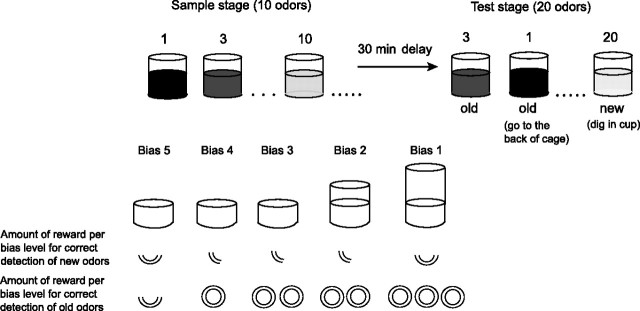
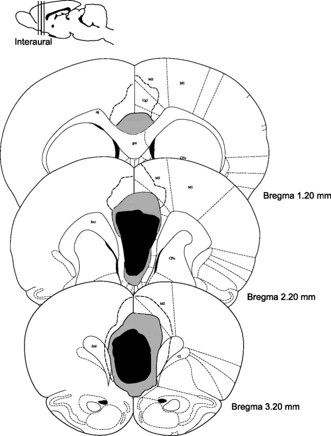
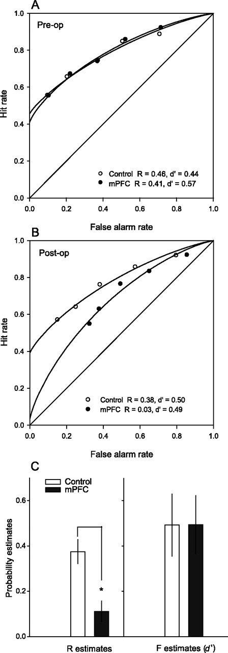
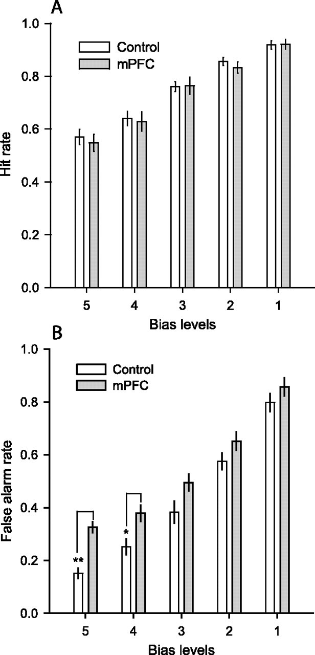
There is continuing controversy about the extent to which the rodent medial prefrontal cortical area (mPFC) is functionally homologous to the dorsolateral prefrontal cortex in humans and nonhuman primates. Previous studies have compared the effects of mPFC lesions in rats to those of dorsolateral prefrontal lesions in working memory, strategy switching, and temporal ordering. None, however, has examined the role of the rodent mPFC in recognition memory, wherein, in humans, dorsolateral prefrontal damage results in a deficit in source monitoring resulting in impaired recollection. In the present study, we examined recognition memory in rats with bilateral mPFC lesions (prelimbic/infralimbic regions; ibotenic acid) using a variant of a non-match-to-sample task with manipulations of response bias that allowed for a signal detection analysis that distinguishes recollection and familiarity contributions to recognition memory. Animals with medial prefrontal lesions had a modest overall deficit in recognition with no general change in their tendency to elicit "old" or "new" responses. Signal detection analyses indicated that rats with mPFC damage had a curvilinear and symmetrical receiver operating characteristic (ROC) curve, compared with a curvilinear and asymmetrical ROC curve in control subjects, indicating that mPFC damage severely reduced recollection-based performance, while sparing familiarity. The recollection failure was associated with an impaired ability to reject new items (increased false alarm rate), whereas the identification of old items (hit rate) was normal. This pattern of findings is similar to that observed in humans with dorsolateral prefrontal damage and is complementary to the selective deficit in hit rate observed after hippocampal damage.
Figures
References
-
- Alexander MP, Stuss DT, Fansabedian N. California verbal learning test: performance by patients with focal frontal and non-frontal lesions. Brain. 2003;126:1493–1503. - PubMed
-
- Brown MW, Aggleton JP. Recognition memory: what are the roles of the perirhinal cortex and hippocampus? Nat Rev Neurosci. 2001;2:51–61. - PubMed
-
- Brown VJ, Bowman EM. Rodents models of prefrontal cortical function. Trends Neurosci. 2002;25:340–343. - PubMed
-
- Buckner RL, Kelley WM, Petersen SE. Frontal cortex contributes to human memory formation. Nat Neurosci. 1999;2:311–314. - PubMed
Publication types
MeSH terms
Grants and funding
LinkOut - more resources
Full Text Sources
Other Literature Sources