. 2008 Dec 10;28(50):13621-8.
doi: 10.1523/JNEUROSCI.4785-08.2008.

A critical window for experience-dependent plasticity at whisker sensory relay synapse in the thalamus

Affiliations

A critical window for experience-dependent plasticity at whisker sensory relay synapse in the thalamus

Hao Wang et al. J Neurosci. .

Abstract

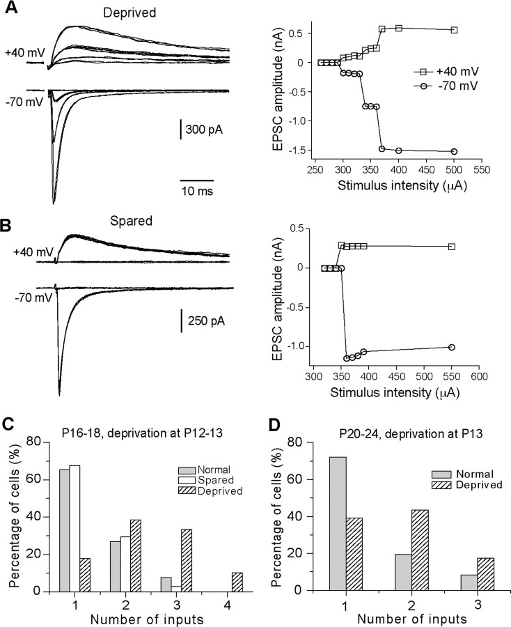
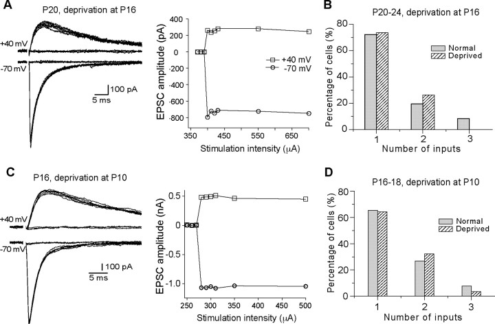
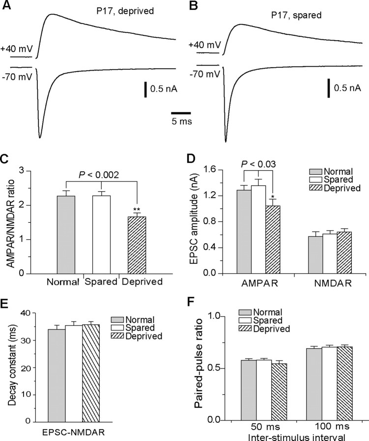
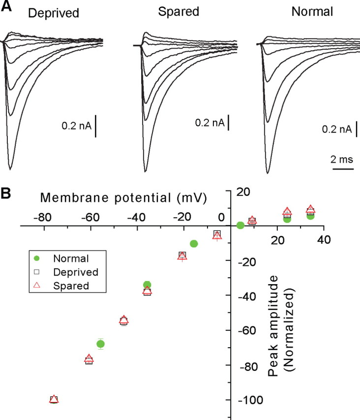
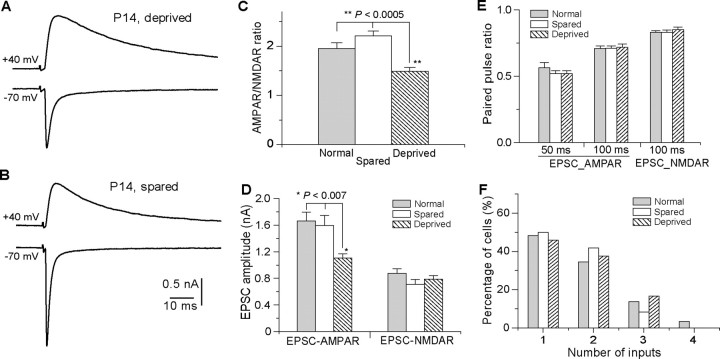
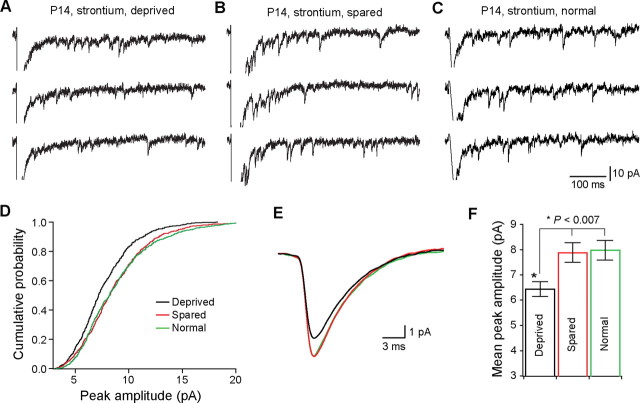
This study investigated the role of sensory experience in the refinement of whisker sensory relay synapses in the ventral posterior medial nucleus (VPm) of the murine thalamus. Sensory deprivation was done by whisker plucking, and synaptic connectivity was determined by whole-cell patch-clamp recording in acute slices. Sensory deprivation started at P12-P13, but not at P16, disrupted the elimination of VPm relay synapses. The majority of deprived neurons received multiple relay inputs, whereas the majority of nondeprived neurons received a single relay input. Sensory deprivation started a few days earlier at P10, however, had no effect on synapse elimination. The disruption of synapse elimination was associated with a delay in synapse maturation. The AMPA/NMDA ratio of EPSC was significantly smaller in deprived neurons. On the other hand, deprivation had no effect on the peak amplitude or decay time constant of the NMDA component, or the I-V relationship of the AMPA component, nor does it affect the paired-pulse ratio of EPSCs. The reduction in the AMPA/NMDA ratio was already evident within 24 h of whisker plucking, and the effect is associated with a reduction in the amplitude of quantal AMPA events. Thus, P12-P13 is a critical period for experience-dependent refinement at the whisker sensory relay synapse in the VPm.


Figures

Figure 1.
Figure 1.
Whisker deprivation started at P12–P13 disrupts synapse elimination at the lemniscal relay connection in the VPm. A, B, Left panels, Synaptic currents in response to stimuli with a range of intensity in VPm neurons at P17 following one-side whisker deprivation started at P13. A is from a neuron in deprived VPm, and B is from a neuron in spared VPm. The right panels are the plots of peak amplitudes at +40 (empty square) or −70 mV (empty circles) versus stimulus intensity. C, The distributions of neurons at P16–P18 with different number of lemniscal inputs for deprived VPm (hatched; n = 39), spared VPm (empty; n = 34), and VPm in normal mice (gray; n = 26). One-side whisker deprivation was performed at P12–P13. D, The distribution of neurons in deprived VPm (hatched; n = 23) at P20–P24 following whisker deprivation started at P13 and the distribution obtained from normal mice at P20–P24 (gray; n = 36).
Figure 2.
Figure 2.
A critical window for the effect of whisker deprivation on synapse elimination. A, Left panel, Synaptic currents at different stimulus intensity recorded from a neuron at P20 following whisker deprivation at P16; right panel, plot of peak amplitude versus stimulus intensity. B, The distribution of neurons (n = 19) in deprived VPm at P20–P24 following whisker deprivation started at P16 and that from normal mice at P20–P24 (in gray; the same data as in Fig. 1D). C, Synaptic responses and the plot of current versus intensity obtained from a neuron at P16 following whisker deprivation at P10. D, The distribution of neurons in deprived VPm (hatched; n = 28) at P16–P18 following whisker deprivation started at P10 and that from normal mice at P16–P18 (gray; the same data as in Fig. 1C).
Figure 3.
Figure 3.
Whisker deprivation started at P13 disrupts maturation of the VPm relay synapse. A, B, Synaptic currents recorded from two neurons in the deprived (A) and spared (B) VPm at P17 following whisker deprivation started at P13. For each cell, synaptic currents at +40 and −70 mV were obtained at the same stimulation intensity. C, The AMPAR/NMDAR ratio obtained from neurons at P16–P18 following whisker deprivation at P13. The ratio obtained from deprived VPm neurons (n = 24; hatched) was significantly smaller than that from spared neurons (n = 27; empty) or neurons in normal mice at the same age (n = 23; gray) (**p < 0.002, Kruskal–Wallis test). D, The peak amplitude of the maximal EPSC-AMPAR and EPSC-NMDAR for deprived (hatched), spared (empty), and normal (gray) VPm neurons at P16–P18 (*p < 0.03, Kruskal–Wallis test). E, Decay constant of EPSC-NMDAR for deprived (hatched), spared (empty), and normal (gray) VPm neurons at P16–P18. F, Paired-pulse ratios of EPSC-AMPAR at interstimulus intervals of 50 and 100 ms.
Figure 4.
Figure 4.
Whisker deprivation does not alter the I–V relationship of EPSC-AMPAR. A, EPSC-AMPAR recorded at various holding potentials from deprived (left), spared (middle), and normal (right) neuron recorded in the presence of d-APV (50 μm). For A (left and middle), the holding potentials, from the top to bottom, were +34, +24, +9, −6, −21, −36, −46, and −76 mV; for A (right), the holding potentials were +34, +24, +4, −16, −36, −56, and −76 mV. B, I–V plots for deprived (black square, n = 7 cells), spared (red triangle, n = 7), and normal (green filled circle, n = 6) groups. For each cell, the peak amplitude was normalized to that recorded at −76 mV. The liquid junction potential, estimated to be +6 mV, was corrected for all cells.
Figure 5.
Figure 5.
Rapid modifications of synaptic properties by whisker deprivation. A and B are evoked synaptic currents from neurons in the deprived (A) and spared (B) VPm of a P14 mouse following one-side whisker deprivation at P13. C, The AMPAR-NMDAR ratio for spared (empty; n = 17), deprived (hatched; n = 19), and normal (gray; n = 20) VPm neurons following acute deprivation (**p < 0.0005, Kruskal–Wallis test). D, Peak amplitudes of EPSC-AMPAR and EPSC-NMDAR in deprived (hatched; n = 24), spared (empty; n = 21), and normal (gray; n = 20) VPm neurons at P14 (*p < 0.007, Kruskal–Wallis test). E, Paired-pulse ratios at 100 ms interval for EPSC-AMPAR and EPSC-NMDAR in deprived (hatched; n = 19), spared (empty; n = 17), and normal (gray; n = 20) VPm neurons at P14. F, Distributions of neurons at P14 with different number of inputs for deprived, spared, and normal groups.
Figure 6.
Figure 6.
Rapid change of quantal events after whisker deprivation. A, B, evoked quantal EPSCs from deprived (A) and spared (B) neuron at P14 following whisker deprivation at P13. C, Evoked quantal EPSCs in a neuron from a normal untreated mouse at P14. Evoked quantal EPSCs (Sr-EPSCs) were recorded at −70 mV in the presence of 3 mm Sr2+ and 0 mm Ca2+. D, Cumulative distributions of the peak amplitude for deprived (black; n = 15 cells), spared (red; n = 15), and normal (green; n = 11) group. For each group, the distribution was established using 50 consecutively detected events from each cell. E, Averaged Sr-EPSCs for deprived (black; n = 15 cells), spared (red; n = 15), and normal (green; n = 11) groups. F, Mean peak amplitudes of Sr-EPSC recorded from deprived, spared, and normal groups (*p < 0.007, Kruskal–Wallis test).

Similar articles

Cited by

References

    1. Allen CB, Celikel T, Feldman DE. Long-term depression induced by sensory deprivation during cortical map plasticity in vivo. Nat Neurosci. 2003;6:291–299. - PubMed
    1. Arsenault D, Zhang ZW. Developmental remodelling of the lemniscal synapse in the ventral basal thalamus of the mouse. J Physiol. 2006;573:121–132. - PMC - PubMed
    1. Belford GR, Killackey HP. The development of vibrissae representation in subcortical trigeminal centers of the neonatal rat. J Comp Neurol. 1979;188:63–74. - PubMed
    1. Bender KJ, Allen CB, Bender VA, Feldman DE. Synaptic basis for whisker deprivation-induced synaptic depression in rat somatosensory cortex. J Neurosci. 2006;26:4155–4165. - PMC - PubMed
    1. Brecht M, Sakmann B. Whisker maps of neuronal subclasses of the rat ventral posterior medial thalamus, identified by whole-cell voltage recording and morphological reconstruction. J Physiol. 2002;538:495–515. - PMC - PubMed

Substances

LinkOut - more resources