. 2008 Dec 10;28(50):13673-83.
doi: 10.1523/JNEUROSCI.4695-08.2008.

SynGAP regulates steady-state and activity-dependent phosphorylation of cofilin

Affiliations

SynGAP regulates steady-state and activity-dependent phosphorylation of cofilin

Holly J Carlisle et al. J Neurosci. .

Abstract

SynGAP, a prominent Ras/Rap GTPase-activating protein in the postsynaptic density, regulates the timing of spine formation and trafficking of glutamate receptors in cultured neurons. However, the molecular mechanisms by which it does this are unknown. Here, we show that synGAP is a key regulator of spine morphology in adult mice. Heterozygous deletion of synGAP was sufficient to cause an excess of mushroom spines in adult brains, indicating that synGAP is involved in steady-state regulation of actin in mature spines. Both Ras- and Rac-GTP levels were elevated in forebrains from adult synGAP(+/-) mice. Rac is a well known regulator of actin polymerization and spine morphology. The steady-state level of phosphorylation of cofilin was also elevated in synGAP(+/-) mice. Cofilin, an F-actin severing protein that is inactivated by phosphorylation, is a downstream target of a pathway regulated by Rac. We show that transient regulation of cofilin by treatment with NMDA is also disrupted in synGAP mutant neurons. Treatment of wild-type neurons with 25 mum NMDA triggered transient dephosphorylation and activation of cofilin within 15 s. In contrast, neurons cultured from mice with a homozygous or heterozygous deletion of synGAP lacked the transient regulation by the NMDA receptor. Depression of EPSPs induced by a similar treatment of hippocampal slices with NMDA was disrupted in slices from synGAP(+/-) mice. Our data show that synGAP mediates a rate-limiting step in steady-state regulation of spine morphology and in transient NMDA-receptor-dependent regulation of the spine cytoskeleton.


Figures
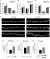
Figure 1.
Figure 1.
Heterozygous synGAP deletion increases the number of mushroom spines on adult hippocampal CA1 neurons in vivo, increases steady-state activation of Ras and Rac, and decreases steady-state activation of cofilin. A, The data show a significant increase (19%) in the number of mushroom-shaped spines per micrometer of dendrite on synGAP+/− hippocampal neurons compared with wild type (wt) [wt = 1.06 ± 0.07 spines/μm dendrite; heterozygous (het) = 1.27 ± 0.05 spines/μm dendrite; p = 0.038; one-tailed Student's t test; n = 3 wt, 3 het mice]. There is no difference in the density of stubby (wt = 0.799 ± 0.034 spines/μm dendrite; het = 0.782 ± 0.030 spines/μm dendrite) or thin spines (wt = 0.407 ± 0.009 spines/μm dendrite; het = 0.387 ± 0.022 spines/μm dendrite). B, The volume of synGAP+/− mushroom spines is significantly larger than wild-type mushroom spines (wt mushroom = 0.070 ± 0.001 μm3, het mushroom = 0.079 ± 0.005 μm3, p = 0.002, Mann–Whitney Rank Sum Test, n = 2383 wt, 2788 het mushroom spines; wt stubby = 0.056 ± 0.002 μm3; het stubby = 0.057 ± 0.003 μm3; wt thin = 0.106 ± 0.012 μm3; het thin = 0.108 ± 0.011 μm3). C, SynGAP+/− mushroom spines were also significantly longer than wild-type mushroom spines (wt mushroom = 0.797 ± 0.029 μm, het mushroom = 0.849 ± 0.034 μm, p = 0.001, Mann–Whitney Rank Sum Test, n = 2383 wt, 2788 het mushroom spines; wt stubby = 0.187 ± 0.002 μm, het stubby = 0.179 ± 0.001 μm; wt thin = 0.622 ± 0.022 μm, het thin = 0.639 ± 0.025 μm). D, Example dendrites are shown from synGAP+/+/GFP+/− and synGAP+/−/GFP+/− mice. E, The level of Ras-GTP in synGAP+/− forebrains was approximately twofold greater than in wild type [wt = 0.49 ± 0.04, het = 0.95 ± 0.11 arbitrary units (a.u.); p = 0.016, two-tailed t test; n = 3 wt, 3 het mice]. F, The level of Rac-GTP in synGAP+/− forebrains was ∼30% higher than in wild type (wt = 2.15 ± 0.46, het = 2.58 ± 0.41 a.u; p = 0.005, paired two-tailed t test; n = 7 wt, 7 het mice). G, Steady-state phosphorylation of cofilin was ∼75% higher in synGAP+/− crude forebrain homogenate than in wild type (wt = 0.779 ± 0.169, het = 1.38 ± 0.138, p = 0.0122, paired two-tailed t test, n = 5 wt, 5 het mice). Bottom in E, F, and G show example blots. Error bars represent SEM.
Figure 2.
Figure 2.
SynGAP is phosphorylated by CaMKII in response to NMDA receptor activation. Experiments were performed on wild-type cortical or hippocampal neuronal cultures after 15–18 DIV. A, SynGAP is basally phosphorylated on Ser1123 in untreated wild-type cortical cultures. The phosphorylation is abolished when cultures are incubated in calcium-free medium (-[Ca2+]e), the NMDAR open-channel blocker MK801 (10 μm), or the calmodulin antagonist W-7 (100 μm) for 30 min (9.7, 13.7, and 0.2% of control, respectively). B, Basal phosphorylation of both synGAP and CaMKII are abolished by a 30 min incubation in 1 μm K252a, a general calmodulin-dependent protein kinase inhibitor (3.5 and 1.9% of control, respectively), and reduced by incubation with 5 μm KN-93, a specific inhibitor of CaMKII (39.6 and 30.8% of control, respectively). Incubation in 10 μm KN-93 did not further reduce the level of phosphorylation (38.2% of control p-SynGAP and 30.8% of control p-CaMKII). C, Regulation of phosphorylation of synGAP (ser-1123) by activation of NMDA receptors was identical in hippocampal and cortical cultures. A 15 s application of 25 μm NMDA or 250 μm NMDA stimulated phosphorylation of synGAP above control levels (25 μm = 188% and 250 μm = 364% of control hippocampal phospho-synGAP; 25 μm = 243% and 250 μm = 398% of control cortical phospho-synGAP). D, In cortical cultures, phosphorylation of CaMKII and synGAP stimulated by a 15 s application of NMDA (25 μm) was blocked by the CaMKII inhibitor KN-93 (10 μm) (NMDA alone = 355% of control phospho-synGAP and 576% of control phospho-CaMKII; NMDA + KN-93 = 151% of control phospho-synGAP and 177% of control phospho-CaMKII). Experiments were performed three times with similar results, except for those in part D, which were performed twice with similar results.
Figure 3.
Figure 3.
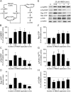
Bath application of NMDA in neuronal cultures increases the phosphorylation of CaMKII and synGAP, the activation of Ras and Rac, and the phosphorylation of PAK, but causes a decrease in the phosphorylation of cofilin. A, The diagram depicts one postulated pathway through which the NMDA receptor regulates Ras activity (left) and the Rac-dependent pathway that regulates cofilin (right). The dotted line represents a hypothetical link between activated Ras and activated Rac that is suggested by our data. Wild-type cortical or hippocampal neurons were treated with NMDA (25 μm) for 0.25, 1, 3, or 5 min. Phosphorylation of CaMKII, synGAP, PAK, and cofilin, and activation of Ras and Rac were measured as described in Materials and Methods. B, E, Samples from cortical and hippocampal cultures that were immunoblotted with anti-phospho-Thr286 CaMKII antibody showed a significant increase in phosphorylation within 15 s of treatment with NMDA (15 s* = 209 ± 23%, p = 0.009; 1 min* = 225 ± 35%, p = 0.016; 3 min* = 212 ± 35%, p = 0.023; 5 min* = 167 ± 25% of untreated control, p = 0.046; one-sample t test, data from cortical and hippocampal cultures were combined, n = 6 independent experiments). C, E, Samples from cortical and hippocampal cultures immunoblotted with anti-phospho-Ser1123 synGAP antibody showed a significant increase in phosphorylation of ser-1123 on synGAP by CaMKII after 15 s, followed by rapid dephosphorylation (15 s* = 189 ± 28%, p = 0.01; 1 min = 146 ± 24%; 3 min = 104 ± 24%; 5 min* = 38.1 ± 16% of untreated control, p = 0.004; two-tailed one-sample t test; data from cortical and hippocampal cultures were combined, n = 10 independent experiments). D, E, Activated Ras was measured in homogenates of hippocampal and cortical cultures as described under Materials and Methods. Treatment with NMDA (25 μm) induced brief activation of Ras [control = 8.32 ± 2.88 arbitrary units (a.u.); 15 s* = 24.8 ± 8.0 a.u., p = 0.018; 1 min = 12.6 ± 2.4 a.u.; 3 min = 8.31 ± 1.71 a.u.; 5 min = 4.50 ± 0.64 a.u.; repeated measures ANOVA; n = 3 independent experiments]. F, E, Activated Rac measured from hippocampal and cortical cultures is significantly elevated after 1 min treatment with NMDA (25 μm) compared with untreated controls (0.25 min = 109 ± 19.8%; 1 min* = 183 ± 22.3%, p = 0.048, two-tailed one-sample t test; 3 min = 207 ± 58.7%; 5 min = 204 ± 54.3% of control; n = 5). G, E, Anti-phospho-Thr423 PAK was elevated more than twofold in the first 15 s of treatment with NMDA (15 s* = 267 ± 46.4%, p = 0.009; 1 min* = 294 ± 54.7%, p = 0.009; 3 min* = 286 ± 63.7%, p = 0.022; 5 min = 132 ± 67.5%; two-tailed, one sample t test; n = 8 independent experiments). H, E Anti-phospho-Ser3 cofilin was dephosphorylated in response to treatment with NMDA for 15 s to 5 min (15 s* = 56.2 ± 8.68%, p > 0.001; 1 min* = 64.2 ± 9.37%, p = 0.005; 3 min* = 64.8 ± 11.3%, p = 0.001; 5 min* = 78.3 ± 9.46%, p = 0.047; two-tailed, one-sample t test; n = 10 independent experiments). Representative immunoblots are shown in E. D shows means ± SEM B, C, F, G and H show GM ±SEs of the SEGM.
Figure 4.
Figure 4.
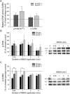
Steady-state activity of PAK and cofilin is altered and regulation cofilin activity by the NMDA receptor is deranged in synGAP mutant hippocampal cultures. Wild-type, synGAP+/−, and synGAP−/− hippocampal cultures (DIV 15–18) were treated with 25 μm NMDA for 15 s to 5 min and then immunoblotted with anti-phospho-Thr423 PAK(1–3) or anti-phospho-Ser3 cofilin antibody. Phosphoprotein levels were measured and quantified as described under methods. A, The levels of phospho-PAK and phospho-cofilin were basally elevated in untreated synGAP−/− hippocampal cultures. Changes in phosphorylation of PAK and cofilin are expressed as a percentage of the wild-type untreated control values (synGAP+/− phospho-PAK = 153 ± 29.2% of wt, n = 7 litters; synGAP−/− phospho-PAK* = 202 ± 43.4% of wt, p = 0.006, n = 5 litters; synGAP+/− phospho-cofilin = 121 ± 18.6% of wt, n = 11 litters; synGAP−/− phospho-cofilin* = 141 ± 12.9% of wt, p = 0.012, n = 10 litters). B, Despite the basal increase in phosphorylated PAK in the mutants, treatment with NMDA still induces an additional increase in phospho-PAK, similar to that in wild types, when each genotype is normalized to its own untreated genotype control (wild type: 15 s = 267 ± 46.4%, 1 min = 294 ± 54.7%, 3 min = 286 ± 63.7%, 5 min = 132 ± 67.5% of wild-type control; n = 8 litters) (SynGAP+/−; 15 s = 316 ± 58.5%, 1 min = 315 ± 58.4%, 3 min = 256 ± 45.7%, 5 min = 134 ± 40.6% of synGAP+/− control; n = 8 litters) (SynGAP−/−; 15 s = 252 ± 40.1%; 1 min = 415 ± 130%; 3 min = 322 ± 95.5%; 5 min = 224 ± 75.7% of synGAP−/− control; n = 8 litters). C, NMDA induces significantly less dephosphorylation of cofilin in synGAP mutant compared with wild-type cultures (wild type: 15 s = 59.7 ± 7.37%; 1 min = 67.4 ± 7.05%; 3 min = 68.7 ± 6.68%; 5 min = 73.4 ± 78.91% of wild-type control; n = 6 litters) (SynGAP+/−: 15 s = 83.5 ± 5.64%; 1 min = 77.8 ± 5.29%; 3 min = 77.2 ± 7.04%; 5 min = 80.4 ± 5.16% of SynGAP+/− control; n = 4 litters) (SynGAP−/−: 15 s* = 96.9 ± 1.07%, p = 0.006; 1 min* = 94.6 ± 4.87%, p = 0.37; 3 min = 81.0 ± 7.48%; 5 min = 78.0 ± 5.85% of SynGAP−/−control; n = 5 litters; one-way ANOVA comparing the three genotypes at each time point, followed by Tukey–Kramer multiple comparison test). All time points were significantly less than 100% in the wild-type neurons; the 1, 3, and 5 min time points were significantly less than 100% in the synGAP+/− neurons; and only the 5 min time point was significantly less than 100% in the synGAP−/− neurons (p < 0.05, two-tailed, one-sample t test). Example blots are shown on right. Graphs represent geometric means ± SEGM.
Figure 5.
Figure 5.
NMDA does not induce long term depression of EPSPs in synGAP+/− hippocampal slices. A, Bath application of NMDA (25 μm, 5 min) induced long-lasting depression of wild-type, but not synGAP+/−, EPSPs. Wild-type ESPSs were significantly depressed at 40 min after NMDA was washed out (71 ± 6% of baseline ESPS slope, p = 0.02, two-tailed, one-sample t test, n = 4 wt mice). EPSPs in synGAP+/− slices were not significantly different from baseline levels 40 min after NMDA washout (101 ± 1% of baseline EPSP slope, n = 5 het mice). Example EPSPs at time = −5 min (1) and time = 40 min (2) are shown above. B, In a similar set of experiments, wild-type and synGAP+/− hippocampal slices were collected for biochemical analysis 2 min before bath application of NMDA (control), immediately after NMDA treatment (0 min), and 5, 10, and 15 min after washout of NMDA (as indicated by arrowheads in A). These slices were homogenized and proteins were immunoblotted with anti-serine3-cofilin antibody and anti-actin antibody as described under Materials and Methods. The level of phosphorylated cofilin was normalized to total actin staining. SynGAP+/− slices had significantly higher levels of phosphorylated cofilin (normalized to control values) than wild types after treatment with NMDA (0 min*, wt = 84.1 ± 9.3%, het = 158 ± 6.9%, p = 0.002; 5 min*, wt = 96.2 ± 10.6%, het = 174 ± 12.4%, p = 0.005; 10 min*, wt = 96.3 ± 7.3%, het = 162 ± 21%, p = 0.019; 15 min*, wt = 94.1 ± 2.1%, het = 194 ± 12.9%; *p = 0.0003, two-tailed t test, n = 4 wt, 3 het mice). C, Representative blots from data reported in graph B. Error bars are geometric means ± SEGM.
Figure 6.
Figure 6.
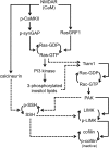
Diagram of regulatory pathways through which synGAP and the NMDA receptor can regulate cofilin activity. Solid lines indicate regulatory interactions that have been shown to occur in spines. Dashed lines indicate regulatory interactions that have been shown to occur in vitro or in non-neuronal cells and that our results suggest also occur in spines. See Discussion for references.

References

    1. Arber S, Barbayannis FA, Hanser H, Schneider C, Stanyon CA, Bernard O, Caroni P. Regulation of actin dynamics through phosphorylation of cofilin by LIM-kinase. Nature. 1998;393:805–809. - PubMed
    1. Bourne J, Harris KM. Do thin spines learn to be mushroom spines that remember? Curr Opin Neurobiol. 2007;17:381–386. - PubMed
    1. Brewer GJ, Torricelli JR, Evege EK, Price PJ. Optimized survival of hippocampal neurons in B27-supplemented Neurobasal, a new serum-free medium combination. J Neurosci Res. 1993;35:567–576. - PubMed
    1. Carlisle HJ, Kennedy MB. Spine architecture and synaptic plasticity. Trends Neurosci. 2005;28:182–187. - PubMed
    1. Chen HJ, Rojas-Soto M, Oguni A, Kennedy MB. A synaptic Ras-GTPase activating protein (p135 SynGAP) inhibited by CaM kinase II. Neuron. 1998;20:895–904. - PubMed

Publication types

MeSH terms

LinkOut - more resources