. 2009 Mar 6;284(10):6436-45.
doi: 10.1074/jbc.M807747200. Epub 2008 Dec 11.

Solution NMR structure of the C-terminal EF-hand domain of human cardiac sodium channel NaV1.5

Affiliations

Solution NMR structure of the C-terminal EF-hand domain of human cardiac sodium channel NaV1.5

Benjamin Chagot et al. J Biol Chem. .

Abstract

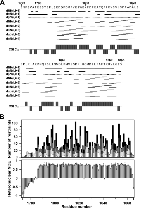
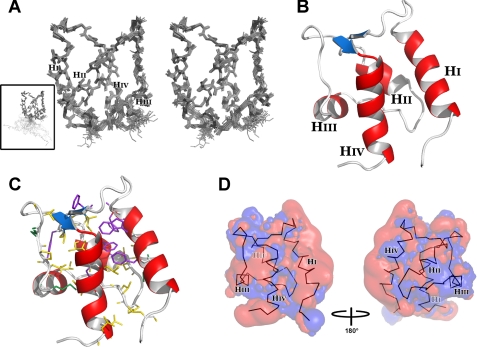
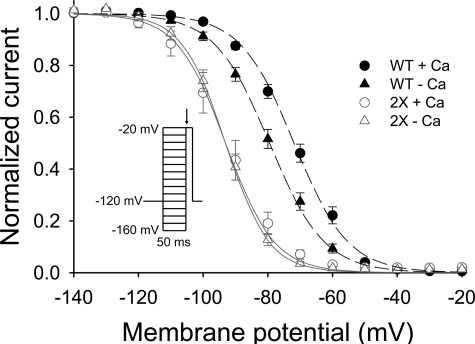
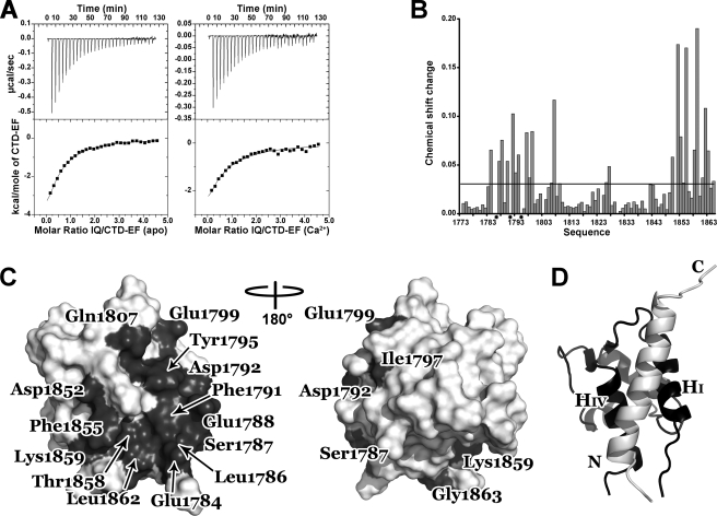
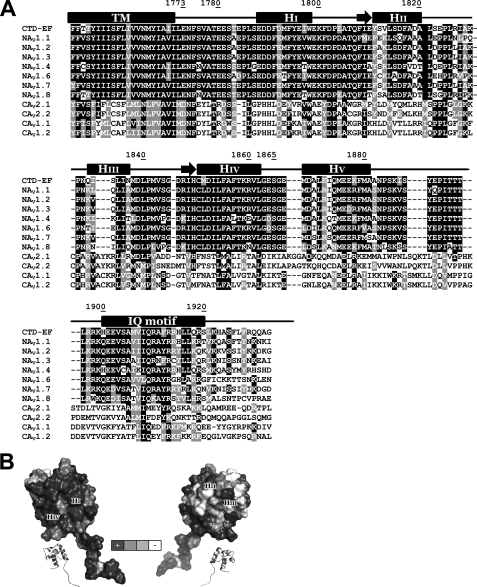
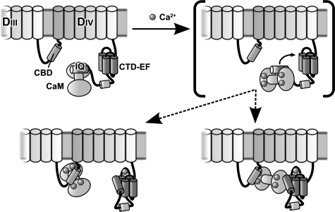
The voltage-gated sodium channel NaV1.5 is responsible for the initial upstroke of the action potential in cardiac tissue. Levels of intracellular calcium modulate inactivation gating of NaV1.5, in part through a C-terminal EF-hand calcium binding domain. The significance of this structure is underscored by the fact that mutations within this domain are associated with specific cardiac arrhythmia syndromes. In an effort to elucidate the molecular basis for calcium regulation of channel function, we have determined the solution structure of the C-terminal EF-hand domain using multidimensional heteronuclear NMR. The structure confirms the existence of the four-helix bundle common to EF-hand domain proteins. However, the location of this domain is shifted with respect to that predicted on the basis of a consensus 12-residue EF-hand calcium binding loop in the sequence. This finding is consistent with the weak calcium affinity reported for the isolated EF-hand domain; high affinity binding is observed only in a construct with an additional 60 residues C-terminal to the EF-hand domain, including the IQ motif that is central to the calcium regulatory apparatus. The binding of an IQ motif peptide to the EF-hand domain was characterized by isothermal titration calorimetry and nuclear magnetic resonance spectroscopy. The peptide binds between helices I and IV in the EF-hand domain, similar to the binding of target peptides to other EF-hand calcium-binding proteins. These results suggest a molecular basis for the coupling of the intrinsic (EF-hand domain) and extrinsic (calmodulin) components of the calcium-sensing apparatus of NaV1.5.


Figures

FIGURE 1.
FIGURE 1.
Distribution of NOEs and correlation with heteronuclear relaxation for the CTD-EF construct. A, distribution of sequential and medium-range NOEs. The NOEs were classified into strong, medium, and weak as indicated by thick, medium, and thin lines, respectively. Chemical shift index values for C-α atoms are indicated. B, histogram of all NOEs (top) and 15N NOEs (bottom). Intraresidue NOEs are in white, sequential NOEs are in light gray, medium-range NOEs are in dark gray, and long-range NOEs are in black. Prolines residues are indicated by stars.
FIGURE 2.
FIGURE 2.
Three-dimensional solution structure of CTD-EF. A, stereo-view of the final ensemble of 20 conformers representing the solution structure, depicted with all backbone atoms. Inset, full-length CTD-EF. B, ribbon diagram with helices labeled and colored red and the cross-strand β-type interaction in blue. C, residues that contribute to the hydrophobic core with aromatic side chains in purple and aliphatic side chains in yellow and green. CTD-EF has the same orientation as in B. D, electrostatic field potentials of CTD-EF with negative charge in red and positive charge in blue. The backbone of CTD-EF is shown within the surface. The four helices are labeled. Contour levels for positive and negative isosurfaces were set to 1 kT and -2 kT, respectively.
FIGURE 3.
FIGURE 3.
Function of NaV1.5 is perturbed by mutation in Ca2+ binding Loop I of CTD-EF. Patch clamp experiments showing the availability curve of the wild-type and D1802A,E1804A mutant (2X) in the absence or presence of Ca2+. Voltage dependence of inactivation is shown for cells expressing wild-type (WT) or 2X Nav 1.5 in high [Ca2+]i (1 mm) or a nominally [Ca2+]i-free solution (20 mm BAPTA). The V½ values for wild-type NaV1.5 were -71.8 ± 1.3 mV (Δ; n = 17) in high [Ca2+]i and -79.4 ± 1.3 mV (Δ; n = 16) in low [Ca2+]i (p < 0.001). The V½ values for 2X NaV1.5 were -93.0 ± 2.7 mV (Δ; n = 10) in high [Ca2+]i and -93.1 ± 1.3 mV (Δ; n = 9) in low [Ca2+]i (p = note significant). Inset, voltage clamp protocol.
FIGURE 4.
FIGURE 4.
Characterization of the interaction between CTD-EF and the IQ motif. A, isothermal titration calorimetry of CTD-EF binding to the IQ motif peptide in the absence (left) and presence (right) of Ca2+. The upper panel shows heat release upon injection of CTD-EF into the IQ peptide solution. The lower panel shows integrated isotherms. B, changes in CTD-EF chemical shifts as the IQ peptide is titrated into the solution. The mean value of the chemical shift change is identified by the horizontal line. Stars indicate residues in “intermediate exchange.” C, interaction surface of CTD-EF. Residues with a chemical shift perturbation more than a half standard deviation above the average are colored dark gray and labeled. D, ribbon diagram of the model of CTD-EF (black) in complex with the IQ motif peptide (white). The N and C termini of the IQ motif and Helices I and IV of CTD-EF are labeled.
FIGURE 5.
FIGURE 5.
Conservation of residues in the C terminus of NaV and CaV channels. A, NaV and CaV sequence alignments. Sequences from top to bottom are shown for CTD-EF NaV1.5 (SWISSPROT Q14524), NaV1.1 (SWISSPROT P35498), NaV1.2 (SWISSPROT Q99250), NaV1.3 (SWISSPROT Q9NY46), NaV1.4 (SWISSPROT P35499), NaV1.6 (SWISSPROT Q9UQD0), NaV1.7 (SWISSPROT Q15858), NaV1.8 (SWISSPROT Q9Y5Y9), CaV2.1 (SWISSPROT O00555), CaV2.2 (SWISSPROT Q00975), CaV1.1 (SWISSPROT Q13698), and CaV1.2 (SWISSPROT Q13936). The residue numbers are indicated for CTD-EF along with secondary structure predicted based on sequence analysis and determined experimentally. B, surface representation of CTD-EF with residues shaded according to the extent of conservation in the NaV family with black indicating complete conservation and white indicating non-conservation.
FIGURE 6.
FIGURE 6.
Model for the action of the Ca2+-sensing apparatus in modulating inactivation gating of NaV1. 5. To simplify the diagram, the transmembrane helices are represented as cylinders on top of the cell membrane, and none of interconnecting loops is shown except for the critical linker between domains III and IV, which is known to mediate inactivation gating. The starting state (low Ca2+) is represented in the upper left panel. There are two alternatives for the final state (high Ca2+) shown in the lower panels. A bracket is drawn around the intermediate state in the upper right panel, which is shown to help explain how the apparatus works. When Ca2+ levels are raised, the interaction of CaM with the IQ motif (IQ) is altered (compare left and right upper panels). The binding of CaM to the IQ motif under high Ca2+ is weaker, which increases the availability of the IQ motif to interact with CTD-EF (lower panels). High Ca2+ also activates CaM interaction with other CaM binding domains (CBD) in NaV1.5 such as that in the linker between domains III and IV (lower panels). This may involve either a bridging interaction (lower right panel) or complete release of the IQ domain (lower left panel).

References

    1. Yu, F. H., and Catterall, W. A. (2003) Genome Biology http://genomebiology.com/2003/4/3/207 - PMC - PubMed
    1. Abriel, H., Cabo, C., Wehrens, X. H., Rivolta, I., Motoike, H. K., Memmi, M., Napolitano, C., Priori, S. G., and Kass, R. S. (2001) Circ. Res. 88 740-745 - PubMed
    1. Wehrens, X. H., Abriel, H., Cabo, C., Benhorin, J., and Kass, R. S. (2000) Circulation 102 584-590 - PubMed
    1. Priori, S. G., Napolitano, C., Gasparini, M., Pappone, C., Della Bella, P., Giordano, U., Bloise, R., Giustetto, C., De Nardis, R., Grillo, M., Ronchetti, E., Faggiano, G., and Nastoli, J. (2002) Circulation 105 1342-1347 - PubMed
    1. Vatta, M., Dumaine, R., Antzelevitch, C., Brugada, R., Li, H., Bowles, N. E., Nademanee, K., Brugada, J., Brugada, P., and Towbin, J. A. (2002) Mol. Genet. Metab. 75 317-324 - PubMed

Publication types

MeSH terms

Associated data