. 2009 Feb;37(2):647-57.
doi: 10.1093/nar/gkn1000. Epub 2008 Dec 11.

DNA damage alters DNA polymerase delta to a form that exhibits increased discrimination against modified template bases and mismatched primers

Affiliations

DNA damage alters DNA polymerase delta to a form that exhibits increased discrimination against modified template bases and mismatched primers

Xiao Meng et al. Nucleic Acids Res. 2009 Feb.

Abstract

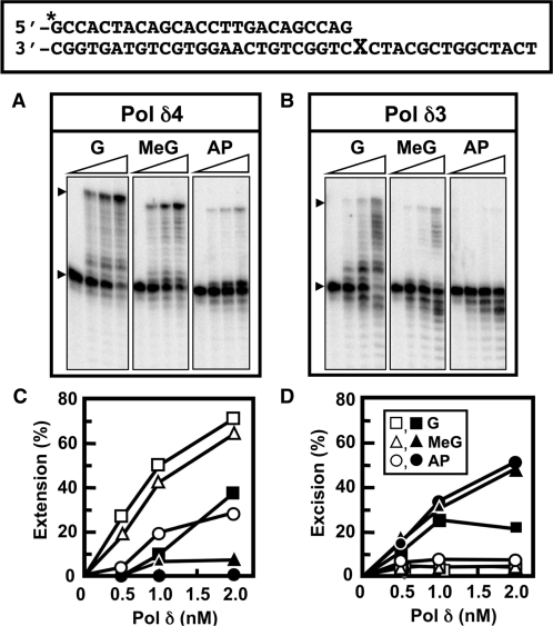
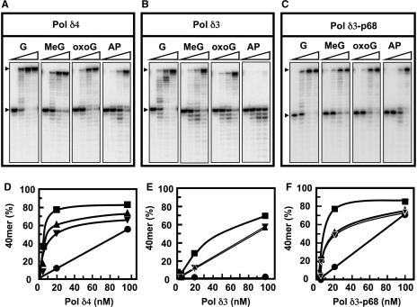
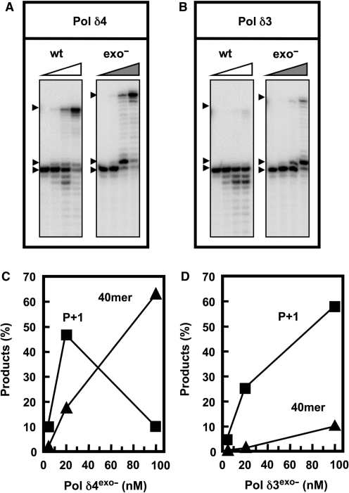
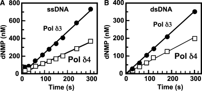
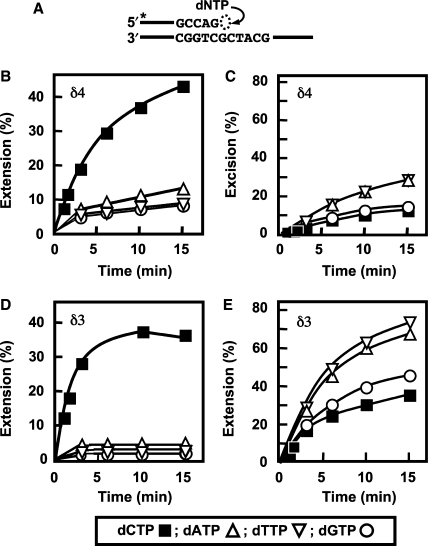
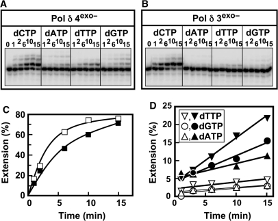
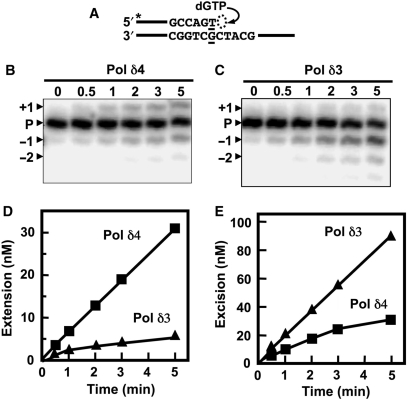
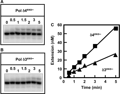
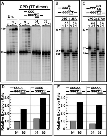
Human DNA polymerase delta (Pol delta4), a key enzyme in chromosomal replication, is a heterotetramer composed of the p125, p50, p68 and p12 subunits. Genotoxic agents such as UV and alkylating chemicals trigger a DNA damage response in which Pol delta4 is converted to a trimer (Pol delta3) by degradation of p12. We show that Pol delta3 has altered enzymatic properties: it is less able to perform translesion synthesis on templates containing base lesions (O(6)-MeG, 8-oxoG, an abasic site or a thymine-thymine dimer); a greater proofreading activity; an increased exonuclease/polymerase activity ratio; a decreased tendency for the insertion of wrong nucleotides, and for the extension of mismatched primers. Overall, our findings indicate that Pol delta3 exhibits an enhanced ability for the detection of errors in both primers and templates over its parent enzyme. These alterations in Pol delta3 show that p12 plays a major role in Pol delta4 catalytic functions, and provides significant insights into the rationale for the conversion of Pol delta4 to Pol delta3 in the cellular response to DNA damage.


Figures

Figure 1.
Figure 1.
Pol δ3 has a reduced ability for the bypass of template lesions when compared to Pol δ4. Pol δ4 and Pol δ3 were prepared by immunoaffinity chromatography from control and UV-treated HeLa cells (see ‘Materials and methods’ section) and assayed for their activities on oligonucleotide primer templates (upper box) containing G, O6-MeG (MeG) or an AP site at position 26 (‘X’). Reactions contained 150 nM 5′-[32P]end-labeled 25-/40-mer primer/template, 400 nM PCNA and 500 μM of each dNTP (see ‘Materials and methods’ section). Products were resolved on 16% denaturing gels and quantitated by phosphorimaging. (A, B) Phosphorimages of the reaction products formed by Pol δ4 and Pol δ3, respectively. For each panel the lanes represent the products formed by 0, 0.5, 1 or 2 nM enzyme. Arrowheads mark the positions of the primer and the full-length 40-mer product. (C, D) Plots of the total amounts of extension products (>25-mer) and excision products (<25-mer), respectively. Products were quantitated and expressed as percentages of the starting primer. Key for symbols [inset, (D)]: template with X = G (squares); O6-MeG (triangles); AP site (circles). Data for Pol δ4 are shown as open symbols and for Pol δ3 as solid symbols.
Figure 2.
Figure 2.
Translesion synthesis by recombinant Pol δ. Recombinant Pol δ4, Pol δ3 and the Pol δ trimer lacking the p68 subunit (Pol δ3-p68) were purified to near-homogeneity (see ‘Materials and methods’ section). Translesion synthesis was assayed using 25-/40-mer templates, where nt 26 = G in the unmodified template, and the modified templates where nt 26 = O6-MeG, 8-oxoG or an AP site under the same conditions as described for Figure 1. Polymerase concentrations used were 0, 4, 20 and 100 nM for the four lanes in each panel. (A–C) Phosphorimages of the reaction products formed by Pol δ4, Pol δ3 and Pol δ3-p68, respectively. (D–F) The amounts of full-length 40-mer products produced by Pol δ4, Pol δ3, and Pol δ3-p68, respectively, were plotted as percentage of the starting 25-mer primer against enzyme concentration. The arrowheads mark the positions of the 40-nt full-length product and the 25-nt primer.
Figure 3.
Figure 3.
Bypass synthesis on AP containing templates by wt and exonuclease-deficient forms of Pol δ. The recombinant Pol δ4 and Pol δ3 enzymes containing the D402A mutants in the p125 subunit were used (see ‘Materials and methods’ section). These exonuclease-deficient forms of Pol δ were assayed for their abilities to bypass the AP lesion as described in Figure 2; enzyme concentrations were 0, 4, 20 and 100 nM. (A, B) Phosphorimages of the reaction products produced by Pol δ4exo– and Pol δ3exo–, respectively. Arrowheads show the position of the 40-mer, the P+1 product (26-mer) and the primer (25-mer). (C, D) The amounts of the P+1 (squares) and full-length extension products (triangles) were plotted against enzyme concentrations for Pol δ4exo– and Pol δ3exo–, respectively.
Figure 4.
Figure 4.
The 3′ to 5′ exonuclease activities of Pol δ4 and Pol δ3. (A, B) Time courses for the exonuclease activities assayed on a ssDNA substrate (5′-[32P]end-labeled 24-nt oligonucleotide), and a 24-/36-mer dsDNA substrate, respectively (see ‘Materials and methods’ section). Data for Pol δ4 are shown as open squares and for Pol δ3 as solid circles. Reactions contained 4 nM polymerases, 100 nM 24-mer ssDNA or 24-/36-mer dsDNA; MgCl2 was added to initiate the reactions. The concentration of dNMP that was released in order to attain the product distribution found was calculated (see ‘Materials and methods’ section). The specific activities (apparent kcat) of the 3′ to 5′ exonuclease activities were determined from the slopes of the curves divided by the enzyme concentration (4 nM); these were 0.53 s–1 and 0.26 s–1, respectively, for Pol δ3 and Pol δ4 activity on ssDNA, and 0.30 s–1 and 0.17 s–1, respectively, on the dsDNA substrate.
Figure 5.
Figure 5.
Comparison of the misinsertion of wrong nucleotides by Pol δ4 and Pol δ3 in the single nucleotide incorporation assay. (A) Diagram of the assay for single nucleotide incorporation. The abilities of Pol δ (5 nM each) to extend the unmodified 25-/40-mer primer/template (200 nM) (Figure 1) in the presence of 400 nM PCNA and 500 μM correct (dCTP) or incorrect nucleotides were examined (see ‘Materials and methods’ section). Reactions were performed for 0, 2, 4, 6, 10 and 15 min. Products were resolved by denaturing gel electrophoresis followed by phosphorimaging and quantitation. (B, C) Extension products and excision products, respectively, for Pol δ4. (D, E) Extension and excision products, respectively, for Pol δ3. Symbols (bottom box): dCTP, solid squares; dTTP, open inverted triangles; dGTP, open circles; dATP, open triangles. Amounts of products were expressed as the percentages of the initial primer amounts.
Figure 6.
Figure 6.
Comparison of the misinsertion of wrong nucleotides by Pol δ4exo– and Pol δ3exo– in the single nucleotide incorporation assay. The exonuclease-deficient forms of Pol δ4 and Pol δ3 were used (see ‘Materials and methods’ section). Assays for single nucleotide incorporation were performed as described in Figure 5. (A, B) Phosphorimages of the gels for the reaction products formed by Pol δ4exo– and Pol δ3exo–, respectively. (C) Quantitation of the amounts of extension products formed expressed as % of the primer converted for the correct nucleotide (dCTP). Data for Pol δ4exo– are shown by the solid squares and data for Pol δ3exo– are shown as open squares. (D) Data for insertion of the wrong nucleotides: dTTP, inverted triangles; dGTP, circles; dATP, triangles. Data for Pol δ4exo– are shown by the solid symbols and data for Pol δ3exo– are shown as open symbols.
Figure 7.
Figure 7.
Comparison of the extension of a mismatched primer terminus by Pol δ4 and Pol δ3 (A) Diagram of the assay for mismatch extension. The abilities of Pol δ4 and Pol δ3 to extend a 26-/40-mer primer/template in which the 3′ nt of the primer forms a T:G mismatch (underlined) with the template was examined. The reactions contained 2 nM Pol δ4 or Pol δ3, 400 nM PCNA, 250 nM 26-/40-mer and 500 μM dGTP. Only the correct next nucleotide (dGTP) was added to the reactions. Reactions were carried out for 0, 0.5, 1, 2, 3 and 5 min. (see ‘Materials and methods’ section). (B, C) Phosphorimages of the reaction products formed by Pol δ4 and Pol δ3, respectively. The positions of the 26-mer primer (‘P’), the 27-mer mismatch extension product (+1), the exonucleolytic 25-mer (–1) and 24-mer (–2) products are shown by the arrowheads. (D, E) The amounts of mismatch extension (27-mer) and excision products (>26mer), respectively, that were formed. Product formation was expressed as concentrations formed in the assay in nanomoles, based on the initial concentration of the primer. Data for Pol δ4 are shown as squares and for Pol δ3 as triangles.
Figure 8.
Figure 8.
Comparison of the extension of a mismatched primer terminus by Pol δ4exo– and Pol δ3exo–. Assays for mismatch extension were performed as in Figure 7. (A, B) Phosphorimages of the reaction products formed by Pol δ4exo– and Pol δ3exo–, respectively. (C) Mismatch extension products (27 nt) formed by Pol δ4exo– and Pol δ3exo– were quantitated and are shown as squares and triangles, respectively.
Figure 9.
Figure 9.
Translesion synthesis by Pol δ4 and Pol δ3 on a template containing a thymine–thymine dimer. The abilities of Pol δ4 and Pol δ3 to extend primers on a template containing a thymine–thymine dimer (CPD) were examined. The template used was a 40-mer containing a thymine–thymine dimer at nts 26 and 27. The primers used were a 25-mer primer, two 26-mer primers that had an added A (‘26A’) or G (‘26G’) at position 26, and two 27-mer primers that had two added As (27AA’) or Gs (‘27GG’) (see ‘Materials and methods’ section). (A) Phosphorimage of the reaction products formed on a template containing a thymine–thymine dimer. DNA polymerases (4, 20 and 100 nM) were incubated with 500 μM dNTPs, 400-nM PCNA, 100-nM 25-mer end-labeled primer annealed to the unmodified (‘Un’) template or with the CPD template as indicated. Reactions were incubated at 37°C for 15 min. The sequence at the primer terminus is shown, where the two double-underlined TT residues represent the thymine–thymine dimer. From left to right: Pol η activity on the unmodified template (‘Un’), followed by Pol η, Pol δ4 and Pol δ3 activities on the CPD template. Lane ‘C’ represents controls in which no enzyme was added. (B) Pol η, Pol δ4 and Pol δ3 were assayed as in A at 20-nM concentration using the 26G or 26A primers annealed to the 40-mer template containing the thymine–thymine dimer as shown above the phosphorimage. Assays were performed for 5 min. (C). Pol η, Pol δ4 or Pol δ3 were assayed as in B using the 27GG or 27AA primers annealed to the 40-mer template containing the thymine–thymine dimer as shown above the phosphorimage. (D) The rate of exonucleolytic cleavage of the 26A and 26G primers by Pol δ4 and Pol δ3 were assayed as in (B). Pol δ4 and Pol δ3 were incubated with the 26A or 26G primer annealed to the thymine–thymine dimer template as in (B), and the reactions carried out for 0, 1, 2, 4, 6 and 10 min. The amounts of the 26-mers remaining at each time point was determined, were plotted against time and fitted into a single exponential decay equation to obtain the rate constant of 26-mer disappearance. The values were normalized to the rate for Pol δ4 activity on the 26A primer. Values for Pol δ4 are shown by the shaded bars and for Pol δ3 by the solid bars. (E). The rate of exonucleolytic cleavage of the 27AA and 27GG primers annealed to the 40-mer template containing a CPD lesion by Pol δ4 and Pol δ3 were assayed and plotted as described in D. The values were normalized to the rate for Pol δ4 activity on the 26A primer.

References

    1. Brutlag D, Kornberg A. Enzymatic synthesis of deoxyribonucleic acid. 36. A proofreading function for the 3′ to 5′ exonuclease activity in deoxyribonucleic acid polymerases. J. Biol. Chem. 1972;247:241–248. - PubMed
    1. Johnson A, O'Donnell M. Cellular DNA replicases: components and dynamics at the replication fork. Annu. Rev. Biochem. 2005;74:283–315. - PubMed
    1. Joyce CM, Steitz TA. Function and structure relationships in DNA polymerases. Annu. Rev. Biochem. 1994;63:777–822. - PubMed
    1. Kunkel TA, Bebenek K. DNA replication fidelity. Annu. Rev. Biochem. 2000;69:497–529. - PubMed
    1. Johnson KA. Conformational coupling in DNA polymerase fidelity. Annu. Rev. Biochem. 1993;62:685–713. - PubMed

Publication types