. 2008 Dec 23;105(51):20263-8.
doi: 10.1073/pnas.0807056106. Epub 2008 Dec 11.

Fluorescence detection of the movement of single KcsA subunits reveals cooperativity

Affiliations

Fluorescence detection of the movement of single KcsA subunits reveals cooperativity

Rikard Blunck et al. Proc Natl Acad Sci U S A. .

Abstract

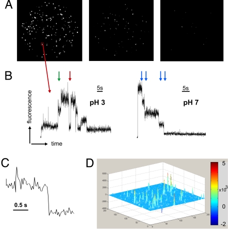
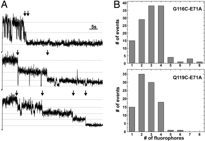
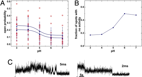
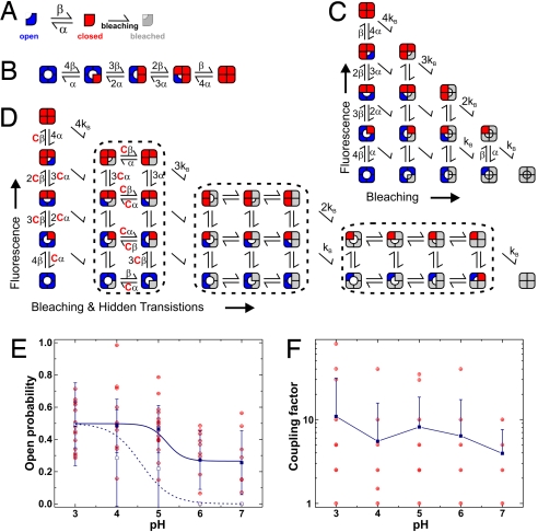
The prokaryotic KcsA channel is gated at the helical bundle crossing by intracellular protons and inactivates at the extracellular selectivity filter. The C-terminal transmembrane helix has to undergo a conformational change for potassium ions to access the central cavity. Whereas a partial opening of the tetrameric channel is suggested to be responsible for subconductance levels of ion channels, including KcsA, a cooperative opening of the 4 subunits is postulated as the final opening step. In this study, we used single-channel fluorescence spectroscopy of KcsA to directly observe the movement of each subunit and the temporal correlation between subunits. Purified KcsA channels labeled at the C terminus near the bundle crossing have been inserted into supported lipid bilayer, and the fluorescence traces analyzed by means of a cooperative or independent Markov model. The analysis revealed that the 4 subunits do not move fully independently but instead showed a certain degree of cooperativity. However, the 4 subunits do not simply open in 1 concerted step.


Conflict of interest statement

The authors declare no conflict of interest.

Figures

Fig. 1.
Fig. 1.
Single-channel imaging of KcsA-Q119C-E71A labeled with TMRM. (A) Distribution of channels in the field of view (Ø ≈ 90 μm). Over time the spots are photobleaching until the last frame (Right) is almost completely bleached. Shown are frames taken with brightfield (BF) at 0.75 s, 24 s, and 60 s. (B) The intensity changes over time are measured within a 3 × 3 pixel square region (red box) around the center of the spot. Shown are 2 time traces at pH 3 and pH 7, respectively. In the left trace, channel activity can be observed. High intensity reflects a closed subunit (4). The subunits may act independently (green arrow) or cooperatively (red arrow). In the right trace (pH 7), no activity was observed. The blue arrows indicate the bleaching steps. (C) Multiple transition step in B shown on an expanded time scale. (D) Three-dimensional representation of the entire image intensity averaged over the first one-third of the series, from which the intensity averaged over the last one-third of the image series is subtracted. The spots appear as strong peaks, indicating that the channels are not diffusing in the supported bilayer (moving spots would disappear in the average and not form peaks). It also indicates that the majority of spots are bleached during the recording (bleached spots are positive in the first third, but zero in the last third).
Fig. 2.
Fig. 2.
Subunit counting. (A) Because of its tetrameric structure, cysteine-modified KcsA can bind up to 4 thiol-reactive fluorophores. We analyzed the time traces with respect to how many bleaching steps are observed. Here, 3 different time traces recorded with TIR at pH 5 are shown with 2, 3, and 4 bleaching steps (arrows). (B) Histograms of G116C-E71A (Upper) and Q119C-E71A (Lower) obtained from counting the bleaching steps from time traces. The probability of a subunits being fluorescent was 0.72 and 0.59 for G116C and Q119C, respectively.
Fig. 3.
Fig. 3.
Open probability determined directly from fluorescence time traces. (A) Open probability of 1 subunit determined from the time traces (BF) considering that the closed subunit is bright and the open subunit is dark. The red spots are single values. Mean and SD are shown in blue. Filled symbols are mean of all values, open symbols are corrected values, which excluded all time traces with no activity (Fig. 3B). P = 0.014/0.006 (ANOVA). (B) pH dependence of fraction of spots showing no activity. (C) Image series (BF) were recorded with higher time resolution (5 and 2 ms, respectively). Whereas the signal-to-noise ratio decreased, no additional information was observed.
Fig. 4.
Fig. 4.
Markov model fitting of KcsA fluorescence time traces. (A) Each subunit may be in the fluorescent (closed, red) or nonfluorescent (open, blue) state. After bleaching, the states fuse into the bleached, nonfluorescent state (gray), where we cannot determine the state of the channel anymore. (B) If the subunits are independent, the rate constants for opening of a single subunit are multiplied by the number of closed subunits and so on. This model indicates the independent, nonbleaching channel. (C) Introducing irreversible bleaching into the model results in a 15-state model, where the bleached subunits (gray) have an undefined state of the bundle crossing. (D) If the subunits are not independent, we have to introduce an additional coupling constant (see text for details) so that the bleached subunits have an influence on the rate constants of the fluorescent subunits. (E) Open probability of a single subunit calculated from the rate constants α and β. Shown are the actual values (red) and mean ± SD (blue filled; P = 0.001 ANOVA). For comparison the channel open probability of KcsA-E71A-Q119C labeled with TMRM is given (blue open symbol). (F) Coupling constant resulting from Markov model fits of the time traces. Shown are single values (red) as well as mean ± SD (blue; neg. SD omitted in logarithmic scale).

References

    1. Doyle DA, et al. The structure of the potassium channel: Molecular basis of K+ conduction and selectivity. Science. 1998;280:69–77. - PubMed
    1. Shimizu H, et al. Global twisting motion of single molecular KcsA potassium channel upon gating. Cell. 2008;132:67–78. - PubMed
    1. Cordero-Morales JF, et al. Molecular determinants of gating at the potassium-channel selectivity filter. Nat Struct Mol Biol. 2006;13:311–318. - PubMed
    1. Blunck R, Cordero-Morales JF, Cuello LG, Perozo E, Bezanilla F. Detection of the opening of the bundle crossing in KcsA with fluorescence lifetime spectroscopy reveals the existence of two gates for ion conduction. J Gen Physiol. 2006;128:569–581. - PMC - PubMed
    1. Thompson AN, Posson DJ, Parsa PV, Nimigean CM. Molecular mechanism of pH sensing in KcsA potassium channels. Proc Natl Acad Sci USA. 2008;105:6900–6905. - PMC - PubMed

Publication types

LinkOut - more resources