. 2008 Dec 12;322(5908):1655-61.
doi: 10.1126/science.1166777.

A competitive inhibitor traps LeuT in an open-to-out conformation

Affiliations

A competitive inhibitor traps LeuT in an open-to-out conformation

Satinder K Singh et al. Science. .

Abstract

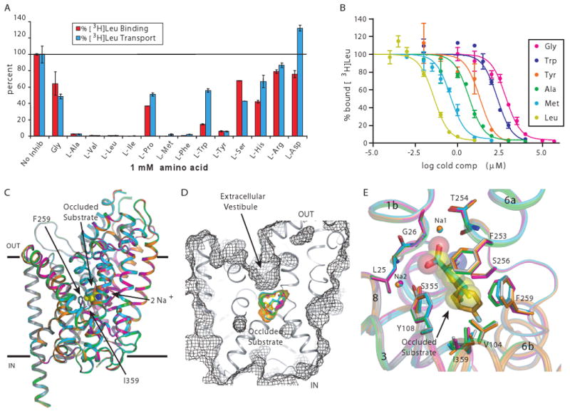
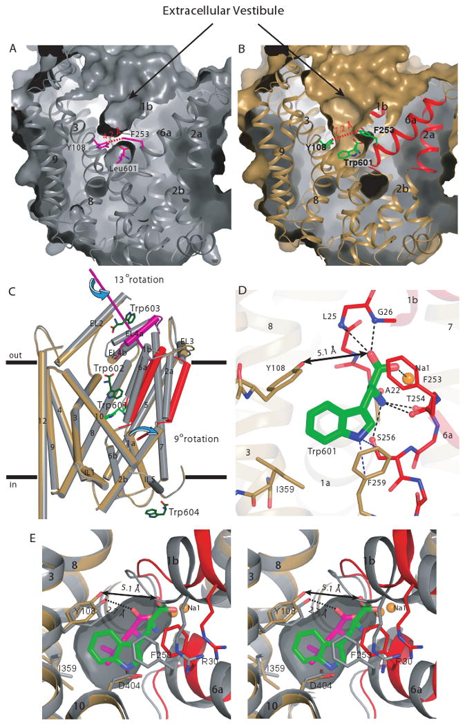
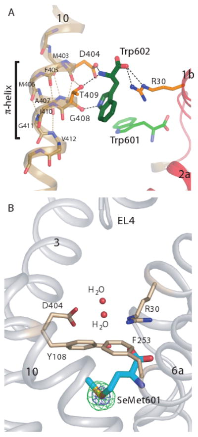
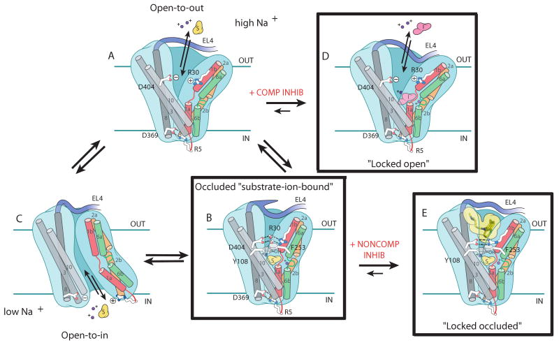
Secondary transporters are workhorses of cellular membranes, catalyzing the movement of small molecules and ions across the bilayer and coupling substrate passage to ion gradients. However, the conformational changes that accompany substrate transport, the mechanism by which a substrate moves through the transporter, and principles of competitive inhibition remain unclear. We used crystallographic and functional studies on the leucine transporter (LeuT), a model for neurotransmitter sodium symporters, to show that various amino acid substrates induce the same occluded conformational state and that a competitive inhibitor, tryptophan (Trp), traps LeuT in an open-to-out conformation. In the Trp complex, the extracellular gate residues arginine 30 and aspartic acid 404 define a second weak binding site for substrates or inhibitors as they permeate from the extracellular solution to the primary substrate site, which demonstrates how residues that participate in gating also mediate permeation.


Figures

Fig. 1
Fig. 1
LeuT substrate screen and occluded state structures. (A) Inhibition of [3H]leucine binding (red bars) and transport (cyan bars) by L-amino acids. (B) Displacement of [3H]leucine binding by leucine (yellow), methionine (cyan), alanine (green), tyrosine (orange), tryptophan (blue), and glycine (magenta). Errors bars represent SEM of triplicate (A) or duplicate (B) measurements. (C) Superposition of the LeuT-Leu (gray), -Ala (green), -Gly (magenta), -Met (cyan), and L-4-F-Phe (orange) complexes using α-carbon positions. Shown in CPK are leucine and the two Na+ ions from the LeuT-Leu complex. Membrane boundaries are demarcated by the two solid black lines. (D) Solvent-accessible surface (depicted in mesh) illustrating the occluded state of the LeuT-substrate complexes. (E) Close-up of the substrate binding pocket, with substrates depicted as sticks. Leucine is shown in semi-transparent CPK representation. Coloring is the same as in (C).
Fig. 2
Fig. 2
Substrate binding pocket - substrate and inhibitor interactions. Substrate binding pockets of the (A) LeuT-Gly, (B) -Ala, (C) -Leu,(D) -Met, (E) -L-4-F-Phe, and (F) -Trp complexes. Hydrogen bonds and polar interactions are illustrated by black dotted lines.
Fig. 3
Fig. 3
Tryptophan is a competitive inhibitor that stabilizes an open-to-outside conformation. Solvent-accessible surface of the (A) LeuT-Leu (gray) and (B) LeuT-Trp complexes (sand/red/magenta). Leucine, tryptophan, Y108, and F253 are depicted in both panels. Distances between Y108 (Cδ1) and F253 (Cξ) in each panel are shown. Helices involved in the domain shift (TM1b, 2a, and 6b) are colored red. (C) Cα superposition (depicted as cylinders) of the LeuT- Leu and LeuT–Trp complexes. Colors are the same as in (A) and (B). EL4a, an additional element involved in the domain shift, is magenta. The rotation axes of the two domains are depicted in their respective colors. The bound tryptophans are shown as stick models, with Trp601 colored bright green and the other three colored dark green. TM11 is omitted from the figure for clarity. (D) Close-up of the Cα superposition depicting the hydrogen bonding network in the substrate binding pocket of the LeuT-Trp complex. Note disruption of the critical hydrogen bond between Y108 and the carboxylate of tryptophan, indicated by a double-headed arrow. (E) Overlay (in stereoview) of the leucine and tryptophan binding sites to illustrate displacement of the ligand α-amino carboxylate group and concomitant shift in protein and sodium positions. Leucine and tryptophan are colored in magenta and green, respectively.
Fig. 4
Fig. 4
A second Trp molecule is bound between R30 and D404 of the extracellular gate only in the open-to-outside conformation. (A) Trp602 bound in the extracellular vestibule of LeuT, residing between D404 and R30, flanked by the π-helix in TM10. (B) Extracellular vestibule of the LeuT-SeMet complex. Anomalous difference Fourier map (contoured at 5σ and 15σ and depicted in green and blue mesh, respectively) showing no significant density peaks in the extracellular vestibule.
Fig. 5
Fig. 5
Schematic of transport and inhibition in LeuT. Postulated conformational changes associated with isomerization from the open-to-out (A) to the outward facing occluded state (B) upon binding of substrate and ions, from the occluded (B) to open-to-in state (C) and dissociation of transported substrate and ions, and from the open-to-in (C) back to the open-to-out state (A). (D) Effect of a competitive inhibitor on transport: stabilizing the open-to-out conformation. (E) TCAs are noncompetitive inhibitors that stabilize the occluded state. The boxed conformations represent actual crystal structures, while the unboxed conformations are hypothetical.

Comment in

References

    1. Parsons SM. FASEB J. 2000;14:2423. - PubMed
    1. Roux MJ, Supplisson S. Neuron. 2000;25:373. - PubMed
    1. Zerangue N, Kavanaugh MP. Nature. 1996;383:634. - PubMed
    1. Chen NH, Reith ME, Quick MW. Pflugers Arch. 2004;447:519. - PubMed
    1. Kanner BI. J Membr Biol. 2006;213:89. - PubMed

Publication types

MeSH terms