. 2009 Feb;191(4):1200-10.
doi: 10.1128/JB.01120-08. Epub 2008 Dec 12.

Synthesis of CDP-activated ribitol for teichoic acid precursors in Streptococcus pneumoniae

Affiliations

Synthesis of CDP-activated ribitol for teichoic acid precursors in Streptococcus pneumoniae

Stefanie Baur et al. J Bacteriol. 2009 Feb.

Abstract

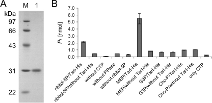
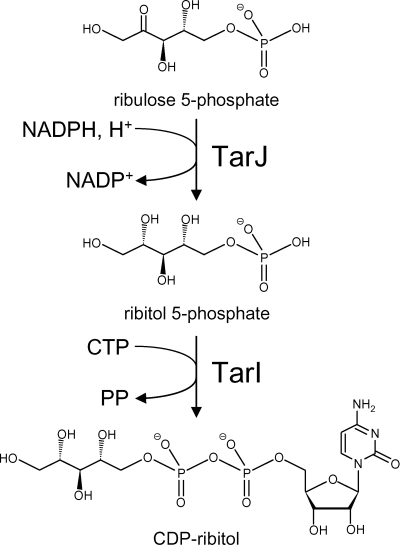
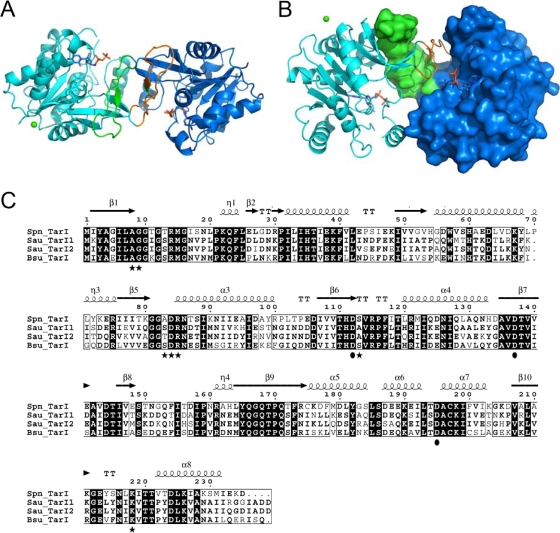
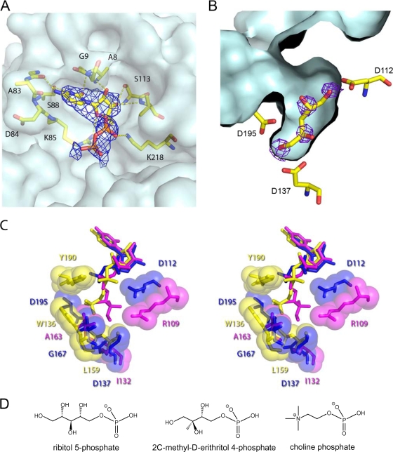
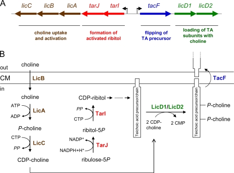
Streptococcus pneumoniae has unusually complex cell wall teichoic acid and lipoteichoic acid, both of which contain a ribitol phosphate moiety. The lic region of the pneumococcal genome contains genes for the uptake and activation of choline, the attachment of phosphorylcholine to teichoic acid precursors, and the transport of these precursors across the cytoplasmic membrane. The role of two other, so far uncharacterized, genes, spr1148 and spr1149, in the lic region was determined. TarJ (spr1148) encodes an NADPH-dependent alcohol dehydrogenase for the synthesis of ribitol 5-phosphate from ribulose 5-phosphate. TarI (spr1149) encodes a cytidylyl transferase for the synthesis of cytidine 5'-diphosphate (CDP)-ribitol from ribitol 5-phosphate and cytidine 5'-triphosphate. We also present the crystal structure of TarI with and without bound CDP, and the structures present a rationale for the substrate specificity of this key enzyme. No transformants were obtained with insertion plasmids designed to interrupt the tarIJ genes, indicating that their function could be essential for cell growth. CDP-activated ribitol is a precursor for the synthesis of pneumococcal teichoic acids and some of the capsular polysaccharides. Thus, all eight genes in the lic region have a role in teichoic acid synthesis.


Figures

FIG. 1.
FIG. 1.
Enzymatic activity of TarJ-His (Spr1148-His). (A) Analysis of TarJ-His by SDS-gel electrophoresis. The protein was separated by SDS-12% PAGE, followed by staining with Coomassie blue. Lane 1, TarJ-His; M, molecular mass marker. The molecular sizes of the marker proteins are indicated on the left side. (B) Photometric assay for the consumption of NADPH or NADH. TarJ-His consumed NADPH in the presence of ribulose 5-phosphate. rel, relative. (C) Analysis of the reaction products by electrospray ionization MS/MS. In the sample with enzyme (lower panel) the expected molecular mass of ribitol 5-phosphate (231 Da; negative ionization mode) was present. The panel shows MS/MS analysis of the 231-Da signal and the assignment of the observed masses to the expected fragmentation products of ribitol 5-phosphate. The control sample without enzyme contained a mass signal of 229 Da (corresponding to ribulose 5-phosphate) which was chosen for fragmentation (upper panel). The measured masses and the proposed structure of the fragmentation products are indicated. The calculated masses are given in brackets. cps, counts per second; amu, atomic mass units.
FIG. 2.
FIG. 2.
Enzymatic activity of TarI-His (Spr1149-His). (A) Analysis of TarI-His by SDS-gel electrophoresis. The protein was separated by SDS-12% PAGE, followed by staining with Coomassie blue. Lane 1, TarI-His; M, molecular size marker. The molecular masses of the marker proteins are indicated on the left side. (B) Assay to detect the release of pyrophosphate by TarI-His. The complete sample (left side) contained Spr1149-His, ribitol 5-phosphate (ribitol-5P), CTP, and pyrophosphatase (PPase), the latter converting pyrophosphate to phosphate, which was detected in the malachite green assay. Either control samples lacked one of the assay components, or the components were tested alone for the presence of phosphate. Other possible substrates (2-l-methyl-d-erythritol [MEP], glycerol 1-phosphate [G3P], and phosphorylcholine [Cho-P]) were also tested with TarI or in the absence of enzyme (controls). TarI-His released pyrophosphate from CTP in the presence of ribitol 5-phosphate and MEP but not glycerol 1-phosphate and phosphorylcholine.
FIG. 3.
FIG. 3.
Reactions of TarI and TarJ with the structures of the substrates and products. PP, pyrophosphate.
FIG. 4.
FIG. 4.
The molecular architecture of TarI. (A) A cartoon view of the structure of TarI; the two monomers are shown in cyan and blue, with the arm regions that mediate dimer formation shown in green and orange. CDP is shown in stick representation, and the crystallographic calcium is shown as a green sphere. (B) An orthogonal view of panel A highlighting the dimerization of the protein and the molecular surface of a monomer. The arm region contributes a number of residues to the active site of the enzyme. (C) Sequence comparison of TarI from S. pneumoniae, S. aureus, and B. subtilis W23. Sequences were aligned using the Multalin program (http://bioinfo.genopole-toulouse.prd.fr/multalin/multalin.html). Black and gray boxes indicate strictly and highly conserved amino acids, respectively. The structural elements of S. pneumoniae TarI determined in this work are shown above the sequence. Residues shown to bind CTP are highlighted as black stars below the sequences, and those that bind to the sugar are shown as black circles. Note that S. aureus has two TarI proteins. Accession numbers are as follows: S. aureus (Sau) N315 TarI, NP_373491; S. aureus N315 TarI′, NP_373487; B. subtilis (Bsu) W23 TarI, CAC86109; S. pneumoniae (Spn) R6 TarI (Spr1149), NP_358742.
FIG. 5.
FIG. 5.
Ligand binding site of TarI. (A) The nucleotide pocket with bound CDP is shown with a composite-omit map rendered at 1σ, to show the presence of nucleotide. Residues involved in polar contacts to the nucleotide are shown in ball and stick representation. The surface of the protein is shown in light blue to highlight the pocket. (B) The ribitol pocket of TarI from the apo structure is shown with FobsFcalc density for crystallographic waters, which correspond almost perfectly to potential sites of hydroxyl groups from a ribitol, which has been modeled by hand into the pocket. The corresponding contact residues are shown in stick representation with the ligand pocket surface shown in light blue. (C) Ligand binding pockets of cytidylyl transferases. Stereo overview of the ligand binding pockets is shown for those structures in the PDB with bound product. TarI (PDB 2VSH) is shown in blue, the MEP cytidylyl transferase IspD (PDB 1INI) is shown in magenta, and phosphorylcholine cytidylyl transferase LicC (PDB 1JYL) is in yellow. Key contact residues are labeled in the corresponding color for the protein. All ligands are shown in stick representation with contact residues shown in stick and sphere representation. (D) Structure of the substrates of cytidylyl transferases.
FIG. 6.
FIG. 6.
Genetic organization of the lic region and functions of the gene products. (A) The lic region contains eight genes with functions in teichoic acid synthesis. The promoters of the lic1 operon (with the tarI, tarJ, licA, licB, and licC genes) and the lic2 operon (with the tacF, licD1, and licD2 genes) are shown as arrows. The functions of the gene products are written below the gene(s). (B) Roles of the gene products in the formation of the teichoic acid precursor and its transport across the membrane. CM, cytoplasmic membrane; P, phosphate residue.

References

    1. Aanensen, D. M., A. Mavroidi, S. D. Bentley, P. R. Reeves, and B. G. Spratt. 2007. Predicted functions and linkage specificities of the products of the Streptococcus pneumoniae capsular biosynthetic loci. J. Bacteriol. 1897856-7876. - PMC - PubMed
    1. Badger, J., J. M. Sauder, J. M. Adams, S. Antonysamy, K. Bain, M. G. Bergseid, S. G. Buchanan, M. D. Buchanan, Y. Batiyenko, J. A. Christopher, S. Emtage, A. Eroshkina, I. Feil, E. B. Furlong, K. S. Gajiwala, X. Gao, D. He, J. Hendle, A. Huber, K. Hoda, P. Kearins, C. Kissinger, B. Laubert, H. A. Lewis, J. Lin, K. Loomis, D. Lorimer, G. Louie, M. Maletic, C. D. Marsh, I. Miller, J. Molinari, H. J. Muller-Dieckmann, J. M. Newman, B. W. Noland, B. Pagarigan, F. Park, T. S. Peat, K. W. Post, S. Radojicic, A. Ramos, R. Romero, M. E. Rutter, W. E. Sanderson, K. D. Schwinn, J. Tresser, J. Winhoven, T. A. Wright, L. Wu, J. Xu, and T. J. Harris. 2005. Structural analysis of a set of proteins resulting from a bacterial genomics project. Proteins 60787-796. - PubMed
    1. Baykov, A. A., O. A. Evtushenko, and S. M. Avaeva. 1988. A malachite green procedure for orthophosphate determination and its use in alkaline phosphatase-based enzyme immunoassay. Anal. Biochem. 171266-270. - PubMed
    1. Bentley, S. D., D. M. Aanensen, A. Mavroidi, D. Saunders, E. Rabbinowitsch, M. Collins, K. Donohoe, D. Harris, L. Murphy, M. A. Quail, G. Samuel, I. C. Skovsted, M. S. Kaltoft, B. Barrell, P. R. Reeves, J. Parkhill, and B. G. Spratt. 2006. Genetic analysis of the capsular biosynthetic locus from all 90 pneumococcal serotypes. PLoS Genet. 2e31. - PMC - PubMed
    1. Bernal, C., C. Palacin, A. Boronat, and S. Imperial. 2005. A colorimetric assay for the determination of 4-diphosphocytidyl-2-C-methyl-d-erythritol 4-phosphate synthase activity. Anal. Biochem. 33755-61. - PubMed

Publication types

LinkOut - more resources