. 2009 Apr;191(7):2012-22.
doi: 10.1128/JB.01547-08. Epub 2008 Dec 12.

Mutations in ampG and lytic transglycosylase genes affect the net release of peptidoglycan monomers from Vibrio fischeri

Affiliations

Mutations in ampG and lytic transglycosylase genes affect the net release of peptidoglycan monomers from Vibrio fischeri

Dawn M Adin et al. J Bacteriol. 2009 Apr.

Abstract

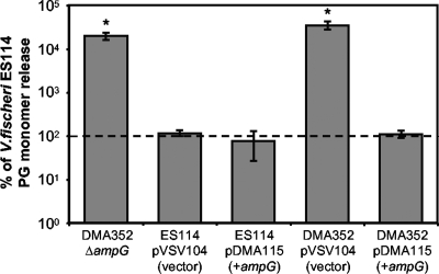
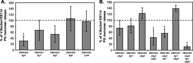
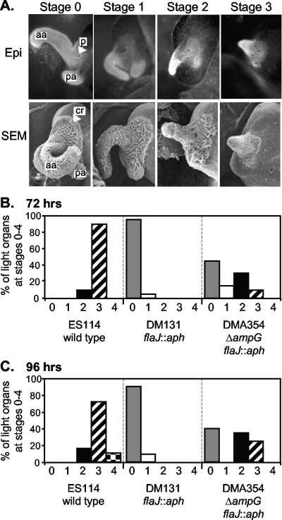
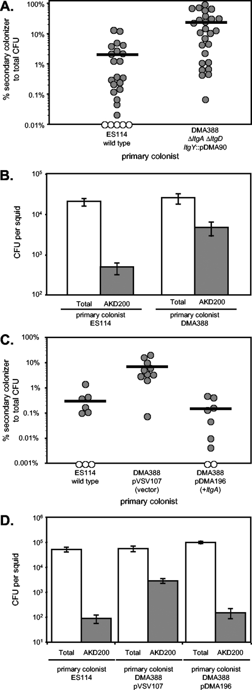
The light-organ symbiont Vibrio fischeri releases N-acetylglucosaminyl-1,6-anhydro-N-acetylmuramylalanyl-gamma-glutamyldiaminopimelylalanine, a disaccharide-tetrapeptide component of peptidoglycan that is referred to here as "PG monomer." In contrast, most gram-negative bacteria recycle PG monomer efficiently, and it does not accumulate extracellularly. PG monomer can stimulate normal light-organ morphogenesis in the host squid Euprymna scolopes, resulting in regression of ciliated appendages similar to that triggered by infection with V. fischeri. We examined whether the net release of PG monomers by V. fischeri resulted from lytic transglycosylase activity or from defects in AmpG, the permease through which PG monomers enter the cytoplasm for recycling. An ampG mutant displayed a 100-fold increase in net PG monomer release, indicating that AmpG is functional. The ampG mutation also conferred the uncharacteristic ability to induce light-organ morphogenesis even when placed in a nonmotile flaJ mutant that cannot infect the light-organ crypts. We targeted five potential lytic transglycosylase genes singly and in specific combinations to assess their role in PG monomer release. Combinations of mutations in ltgA, ltgD, and ltgY decreased net PG monomer release, and a triple mutant lacking all three of these genes had little to no accumulation of PG monomers in culture supernatants. This mutant colonized the host as well as the wild type did; however, the mutant-infected squid were more prone to later superinfection by a second V. fischeri strain. We propose that the lack of PG monomer release by this mutant results in less regression of the infection-promoting ciliated appendages, leading to this propensity for superinfection.


Figures

FIG. 1.
FIG. 1.
Mutation in V. fischeri ampG increases net PG monomer release in log-phase cultures. PG monomer was measured in culture supernatants of ES114 (wild type) and DMA352 (ΔampG) along with both strains containing plasmids pVSV104 (control vector) and pDMA115 (ampG in pVSV104). To account for experiment-to-experiment variations, within each experiment each strain's calculated PG monomer release was expressed as a percentage of ES114 PG monomer release. Values shown are the averages of three different experiments. The dotted line represents ES114 PG monomer release, defined as 100%, which averaged 20.4 nM per OD595. Error bars are standard errors. Asterisks indicate significant differences (P < 0.05) from ES114 as determined by Student's t test.
FIG. 2.
FIG. 2.
Mutations in V. fischeri lytic transglycosylases result in a decrease in net PG monomer release in log-phase cultures. Values are the averages of three different experiments ± standard errors. The dotted line represents ES114 PG monomer release, defined as 100%. Asterisks in both panels indicate significant differences (P < 0.05) from ES114 as determined by Student's t test. (A) Plasmid-insertion mutants with disruptions in ltgA, ltgY, ltgD, ltgE, and lysM. The wild type averaged 28 nM per OD595. (B) Comparison of mutants with combinations of in-frame deletion mutations ΔltgA and ΔltgD and a plasmid-insertion mutation in ltgY. The wild type for this set of experiments averaged 14 nM per OD595.
FIG. 3.
FIG. 3.
A nonmotile ampG mutant triggers regression of the ciliated appendages of the light organ. (A) Light-organ ciliated appendages observed by epifluorescence microscopy (Epi) and SEM to illustrate regression stages. Each image is approximately 100-fold enlarged and shows one lobe of a roughly symmetrical bilobed organ. Labels: aa, anterior appendage; pa, posterior appendage; p, one of three closely grouped pores (indicated only in epifluorescence image); cr, ciliated ridge (indicated only in SEM image). Epifluorescence images highlight epithelial cells in the appendages stained with Cell Tracker orange, and SEM images show the fuzzy appearance of the cilia extending from such epithelial cells. In stage 1 regression, fields of ciliated cells thin out, notably in the ciliated ridge and anterior appendage, but both appendages are still present. In stage 2, the posterior appendage is only a nub and the anterior appendage has shortened. In stage 3, the anterior appendage is a nub and the posterior appendage is absent. Stage 4 is not pictured and represents the complete loss of ciliated appendages, as described by Doino and McFall-Ngai (15). (B and C) Freshly hatched juvenile animals were inoculated with ∼2,500 CFU/ml of either ES114 (wild type), DM131 (flaJ::aph), or DMA354 (ΔampG flaJ::aph), each of which was labeled with the gfp-expressing plasmid pVSV102 to confirm that the nonmotile flaJ mutants did not infect the interior light-organ crypts. Animals were stained at 72 h (B) or 96 h (C) postinoculation and scored blindly for regression stage as illustrated in panel A. The data presented are combined from three experiments scored at 72 h and three scored at 96 h, with similar results among each set of three experiments. Twenty to 21 animals inoculated with each strain were scored at 72 h, and 18 to 21 animals inoculated with each strain were scored at 96 h.
FIG. 4.
FIG. 4.
Animals infected with DMA388 (ΔltgA ΔltgD ltgY::pDMA90) are more susceptible to a secondary infection. Hatchlings were inoculated with either ES114, DMA388 (ΔltgA ΔltgD ltgY::pDMA90), DMA388 pVSV107, or DMA388 pDMA196 (ltgA). After 72 h, animals were inoculated with the secondary strain AKD200 (mini-Tn7 Cmr). At 120 h, animals were homogenized and plated to determine both total CFU and Cmr CFU corresponding to the secondary colonizers. Values in panels A and B are the combined averages of six independent experiments, all of which yielded similar results (total n = 25 for ES114 and n = 26 for DMA388). Values in panels C and D are the combined averages of two independent experiments, which yielded similar results (total n = 9 for ES114, n = 10 for DMA388 pVSV107, and n = 10 for DMA388 pDMA196). (A and C) Percentages of total infection comprised by the secondary colonist, AKD200. Each circle represents a single squid. Open circles represent animals below the limit of detection for Cmr cells. Horizontal lines show the averages of all animals within each treatment (panel A, 2% in ES114-infected animals, and 24% in DMA388-infected animals; panel C, 0.3% in ES114-infected animals, 7.4% in DMA388 pVSV107-infected animals, and 0.2% in DMA388 pDMA196-infected animals). (B and D) Averages of both total CFU (open bars) and CFU from secondary infection by AKD200 (shaded bars). Error bars are standard errors. Student t tests indicated that secondary infection by AKD200 in DMA388-infected animals was significantly greater (P < 0.01) than that in ES114-infected animals in panel B and that secondary infection by AKD200 in DMA388 pVSV107-infected animals was significantly greater (P < 0.05) than that in ES114- or DMA388 pDMA196-infected animals in panel D.

Comment in

References

    1. Adin, D. M., N. J. Phillips, B. W. Gibson, M. A. Apicella, E. G. Ruby, M. J. McFall-Ngai, D. B. Hall, and E. V. Stabb. 2008. Characterization of htrB and msbB mutants of the light organ symbiont Vibrio fischeri. Appl. Environ. Microbiol. 74633-644. - PMC - PubMed
    1. Altschul, S. F., W. Gish, W. Miller, E. W. Myers, and D. J. Lipman. 1990. Basic local alignment search tool. J. Mol. Biol. 215403-410. - PubMed
    1. Boettcher, K. J., and E. G. Ruby. 1990. Depressed light emission by symbiotic Vibrio fischeri of the sepiolid squid Euprymna scolopes. J. Bacteriol. 1723701-3706. - PMC - PubMed
    1. Bose, J. L., U. Kim, W. Bartkowski, R. P. Gunsalus, A. M. Overley, N. L. Lyell, K. L. Visick, and E. V. Stabb. 2007. Bioluminescence in Vibrio fischeri is controlled by the redox-responsive regulator ArcA. Mol. Microbiol. 65538-553. - PubMed
    1. Bose, J. L., C. S. Rosenberg, and E. V. Stabb. 2008. Enhancement of symbiotic colonization and conditional attenuation of growth in culture. Arch. Microbiol. 190169-183. - PMC - PubMed

Publication types

MeSH terms

LinkOut - more resources