. 2009 Feb;191(4):1278-92.
doi: 10.1128/JB.01142-08. Epub 2008 Dec 12.

Leucine-responsive regulatory protein (Lrp) acts as a virulence repressor in Salmonella enterica serovar Typhimurium

Affiliations

Leucine-responsive regulatory protein (Lrp) acts as a virulence repressor in Salmonella enterica serovar Typhimurium

Chang-Ho Baek et al. J Bacteriol. 2009 Feb.

Abstract

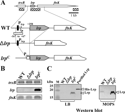
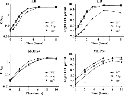
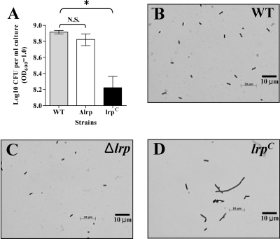
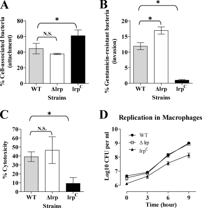
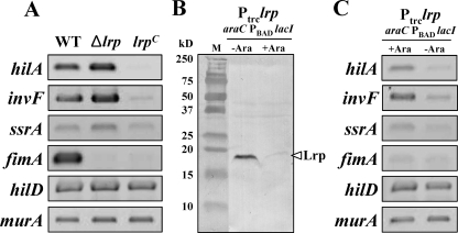
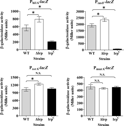
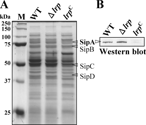
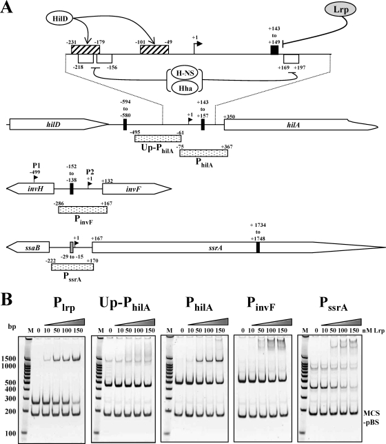
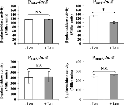
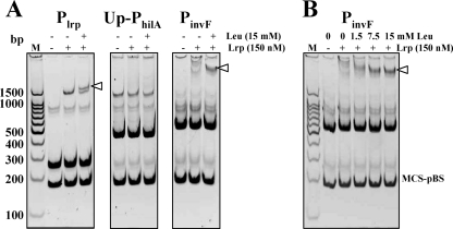
Leucine-responsive regulatory protein (Lrp) is a global gene regulator that influences expression of a large number of genes including virulence-related genes in Escherichia coli and Salmonella. No systematic studies examining the regulation of virulence genes by Lrp have been reported in Salmonella. We report here that constitutive expression of Lrp [lrp(Con)] dramatically attenuates Salmonella virulence while an lrp deletion (Deltalrp) mutation enhances virulence. The lrp(Con) mutant caused pleiotropic effects that include defects in invasion, cytotoxicity, and colonization, whereas the Deltalrp mutant was more proficient at these activities than the wild-type strain. We present evidence that Lrp represses transcription of key virulence regulator genes--hilA, invF, and ssrA--in Salmonella pathogenicity island 1 (SPI-1) and 2 (SPI-2), by binding directly to their promoter regions, P(hilA), P(invF), and P(ssrA). In addition, Western blot analysis showed that the expression of the SPI-1 effector SipA was reduced in the lrp(Con) mutant and enhanced in the Deltalrp mutant. Computational analysis revealed putative Lrp-binding consensus DNA motifs located in P(hilA), P(invF), and P(ssrA). These results suggest that Lrp binds to the consensus motifs and modulates expression of the linked genes. The presence of leucine enhanced Lrp binding to P(invF) in vitro and the addition of leucine to growth medium decreased the level of invF transcription. However, leucine had no effect on expression of hilA and ssrA or on cellular levels of Lrp. In addition, Lrp appears to be an antivirulence gene, since the deletion mutant showed enhanced cell invasion, cytotoxicity, and hypervirulence in BALB/c mice.


Figures

FIG. 1.
FIG. 1.
Analysis of Lrp expression in the wild-type (WT, χ3761), Δlrp (χ9411), and lrp(Con) (χ9448) strains. (A) Schematic diagram of the lrp regions in the Salmonella strains described in the text (hatched box, amplified DNA regions used by RT-PCR; closed boxes, Lrp-binding motif, −79 to −64; dotted box, DNA fragment for EMSA). The coordinates in the picture are numbered with respect to the lrp transcription start site (+1) (66). (B) RT-PCR analysis of the trxB, lrp, and ftsK transcripts from mid-exponential-phase cells grown in LB broth. The images were inverted to intensify the DNA bands. Data are one of two similar RT-PCR results using two independent RNA isolations as a template. (C) Expression of Lrp in the Salmonella strains. Bacterial cells were grown in LB or MOPS minimal medium overnight at 37°C. Whole-cell proteins were resolved on SDS-PAGE (12%) gels. Proteins in the gels were transferred to nitrocellulose for Western blot analysis. Lrp was detected using mouse anti-Lrp serum. Purified His-tagged Lrp protein was used as positive control for the Western blot. Lane M, dual-color prestained protein standards (Bio-Rad).
FIG. 2.
FIG. 2.
Growth curves of the wild-type (WT, χ3761), Δlrp (χ9411), and lrp(Con) (χ9448) strains in LB broth and MOPS minimal broth supplemented with Casamino Acids, vitamins, and trace elements, termed MOPS+. Bacterial cells were grown statically in LB or MOPS+ medium overnight at 37°C and diluted (1:20) in the same medium. Growth was monitored by measuring the OD600 and CFU of the culture at the indicated time intervals. Data are the means ± the standard errors of two independent experiments.
FIG. 3.
FIG. 3.
The lrp(Con) mutation induces elongation of Salmonella cells. (A) Determination of CFU per 1 ml of bacterial cells at an OD600 of 1. Data are means ± standard errors for three separate experiments. *, Values differ significantly from the wild-type strain χ3761 (P < 0.05). N.S., not significant. (B, C, and D) Microscopic comparison of the Salmonella strains grown in LB medium as described in Materials and Methods. The cells were observed and photographed under the oil immersion objective (100× magnification). Data are from one of three independent experiments.
FIG. 4.
FIG. 4.
Analyses of the attachment (A), invasion (B), and cytotoxicity (C) of the wild-type (WT, χ3761), Δlrp (χ9411), and lrp(Con) (χ9448) strains using a mouse IEC line MODE-K and replication of the strains in the macrophagelike cell line J774.A1 (D). Percentages of cell-associated bacteria (attachment) and gentamicin-resistant bacteria (invasion) were calculated with respect to the initial inoculum. The percentages of cytotoxicity were calculated as described in the Cytotox 96 user manual (Promega). To assess bacterial growth in macrophages, the number of bacterial cells remaining after gentamicin treatment was determined. Data are the means ± the standard errors of two separate experiments each performed in duplicate (attachment, and invasion assay) or triplicate (cytotoxicity assay and replication in macrophages). *, Significantly different from the wild-type strain χ3761 (P < 0.05). N.S., not significant.
FIG. 5.
FIG. 5.
RT-PCR analyses of virulence genes in the wild-type (WT, χ3761), Δlrp (χ9411), and lrp(Con) (χ9448) strains. (A and C) RNA was isolated from each strain and used for RT-PCR. The RT-PCR products were separated in a 1.5% agarose gel, stained with ethidium bromide, and visualized on a UV transilluminator. The images were inverted to intensify the DNA bands. Data are one of two similar RT-PCR tests with two independent RNA isolations. (B) Expression of Lrp in strain χ9449 (Ptrc-lrp and araC PBAD lacI) in the presence (+Ara) or absence (−Ara) of arabinose. Lane M, dual-color prestained protein standards (Bio-Rad).
FIG. 6.
FIG. 6.
β-Galactosidase activity in wild-type (WT, χ3761), Δlrp (χ9411), and lrp(Con) (χ9448) strains containing PhilA-lacZ, PinvF-lacZ, PssrA-lacZ, or PmurA-lacZ promoter fusions. Bacterial cells were grown in LB broth to mid-exponential phase and assayed for β-galactosidase activity (69). The PmurA-lacZ fusion was used as the negative control. Data are the means ± the standard errors of two independent experiments. *, Significantly different from the wild-type strain χ3761 (P < 0.05). N.S., not significant.
FIG. 7.
FIG. 7.
Analysis of proteins in culture supernatants of the wild-type (WT, χ3761), Δlrp (χ9411), and lrp(Con) (χ9448) strains. (A) Bacterial cells were grown in LB broth to mid-exponential phase. Cells were removed by centrifugation and proteins in the culture supernatant were precipitated with trichloroacetic acid (10%), resolved in SDS-10% PAGE gel, and stained with Coomassie brilliant blue. (B) Proteins in the gel were subjected to Western blot analysis using anti-SipA rabbit antiserum. Data are one of two similar results from two independent experiments.
FIG. 8.
FIG. 8.
Binding of the purified Lrp to the promoter regions of the virulence-related genes. (A) Depiction of the hilA, invF, and ssrA regions. The stippled boxes represent the PCR fragments used for EMSA. The PCR fragment used for Plrp is shown in Fig. 1A. The binding sites for HilD (hatched box) and H-NS/Hha (open box, AT tract) in PhilA region are shown. The closed boxes and shaded box indicate Lrp-binding consensus II and III, respectively. The coordinates in the picture are numbered with respect to the transcription start sites (+1) (1, 31, 83). There are two transcription start sites, P1 (HilD dependent) and P2 (HilA dependent), in the PinvF region (1). (B) PCR products were incubated with the indicated nanomolar concentrations of Lrp. The reaction mixtures were resolved in a 5% polyacrylamide gel, stained with CYBR Gold (Invitrogen), and visualized on a UV transilluminator. The images were inverted to intensify the DNA bands. Lane M, molecular weight ladder.
FIG. 9.
FIG. 9.
Effect of leucine on expression of hilA, invF, ssrA, and murA. Wild-type Salmonella strains harboring the chromosomal promoter-lacZ fusions, PhilA-lacZ, PinvF-lacZ, PssrA-lacZ, and PmurA-lacZ were grown in MOPS minimal medium for 2 h at 37°C with aeration and split into two portions. Leucine was added into one portion at final concentration of 15 mM (+ Leu), and water (− Leu) was added to another portion of the culture. These cultures were further incubated for 2 h. The bacterial cultures were subjected to assay for β-galactosidase specific activity. Data are the means ± the standard errors of two independent experiments with duplicate. *, Significantly different from the wild-type strain χ3761 (P < 0.05). N.S., not significant.
FIG. 10.
FIG. 10.
Leucine effects on Lrp-DNA interactions. (A) Plrp, Up-PhilA, and PinvF were subjected to EMSA in three different reaction conditions (−/−, without leucine/without Lrp; −/+, without leucine/with Lrp (150 nM); and +/+, with leucine [15 mM]/with Lrp [150 nM]). (B) PinvF-Lrp interaction was analyzed in four different leucine concentrations: 0, 1.5, 7.5, and 15 mM. The binding reaction mixtures were resolved in a 5% polyacrylamide gel, stained with SYBR Gold (Invitrogen), and visualized on a UV transilluminator. The images were inverted to intensify the DNA bands. Data are one of similar results from at least two independent experiments.

References

    1. Akbar, S., L. M. Schechter, C. P. Lostroh, and C. A. Lee. 2003. AraC/XylS family members, HilD and HilC, directly activate virulence gene expression independently of HilA in Salmonella typhimurium. Mol. Microbiol. 47715-728. - PubMed
    1. Altier, C., M. Suyemoto, A. I. Ruiz, K. D. Burnham, and R. Maurer. 2000. Characterization of two novel regulatory genes affecting Salmonella invasion gene expression. Mol. Microbiol. 35635-646. - PubMed
    1. Arricau, N., D. Hermant, H. Waxin, C. Ecobichon, P. S. Duffey, and M. Y. Popoff. 1998. The RcsB-RcsC regulatory system of Salmonella typhi differentially modulates the expression of invasion proteins, flagellin and Vi antigen in response to osmolarity. Mol. Microbiol. 29835-850. - PubMed
    1. Baek, C.-H., and K.-S. Kim. 2003. lacZ- and aph-based reporter vectors for in vivo expression technology. J. Microbiol. Biotechnol. 13872-880.
    1. Baek, C.-H., K.-E. Lee, D.-K. Park, S.-H. Choi, and K.-S. Kim. 2007. Genetic analysis of spontaneous lactose-utilizing mutants from Vibrio vulnificus. J. Microbiol. Biotechnol. 172046-2055. - PubMed

Publication types

MeSH terms

Substances