

Corneal limbal microenvironment can induce transdifferentiation of hair follicle stem cells into corneal epithelial-like cells
- PMID: 19074417
- PMCID: PMC2729676
- DOI: 10.1634/stemcells.2008-0721
Corneal limbal microenvironment can induce transdifferentiation of hair follicle stem cells into corneal epithelial-like cells
Abstract
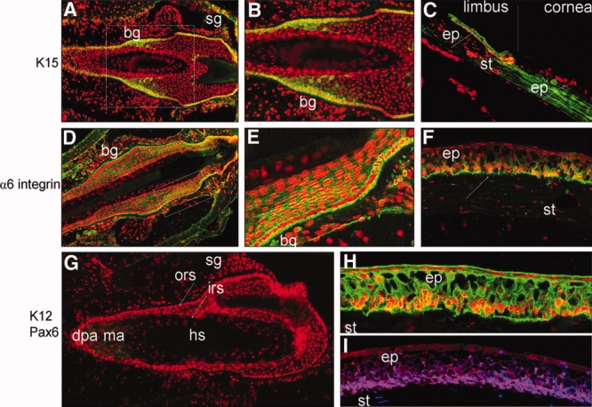
The aim of this study was to investigate the transdifferentiation potential of murine vibrissa hair follicle (HF) stem cells into corneal epithelial-like cells through modulation by corneal- or limbus-specific microenvironmental factors. Adult epithelial stem cells were isolated from the HF bulge region by mechanical dissection or fluorescence-activated cell sorting using antibodies to alpha6 integrin, enriched by clonal expansion, and subcultivated on various extracellular matrices (type IV collagen, laminin-1, laminin-5, fibronectin) and in different conditioned media derived from central and peripheral corneal fibroblasts, limbal stromal fibroblasts, and 3T3 fibroblasts. Cellular phenotype and differentiation were evaluated by light and electron microscopy, real-time reverse transcription-polymerase chain reaction, immunocytochemistry, and Western blotting, using antibodies against putative stem cell markers (K15, alpha6 integrin) and differentiation markers characteristic for corneal epithelium (K12, Pax6) or epidermis (K10). Using laminin-5, a major component of the corneo-limbal basement membrane zone, and conditioned medium from limbal stromal fibroblasts, clonally enriched HF stem and progenitor cells adhered rapidly and formed regularly arranged stratified cell sheets. Conditioned medium derived from limbal fibroblasts markedly upregulated expression of cornea-specific K12 and Pax6 on the mRNA and protein level, whereas expression of the epidermal keratinocyte marker K10 was strongly downregulated. These findings suggest that adult HF epithelial stem cells are capable of differentiating into corneal epithelial-like cells in vitro when exposed to a limbus-specific microenvironment. Therefore, the HF may be an easily accessible alternative therapeutic source of autologous adult stem cells for replacement of the corneal epithelium and restoration of visual function in patients with ocular surface disorders.
Figures


Similar articles
-
From hair to cornea: toward the therapeutic use of hair follicle-derived stem cells in the treatment of limbal stem cell deficiency.Stem Cells. 2011 Jan;29(1):57-66. doi: 10.1002/stem.550. Stem Cells. 2011. PMID: 20957740 Free PMC article.
-
Corneal epithelial-like transdifferentiation of hair follicle stem cells is mediated by pax6 and beta-catenin/Lef-1.Cell Biol Int. 2009 Aug;33(8):861-6. doi: 10.1016/j.cellbi.2009.04.009. Epub 2009 Apr 23. Cell Biol Int. 2009. PMID: 19393751
-
WNT7A and PAX6 define corneal epithelium homeostasis and pathogenesis.Nature. 2014 Jul 17;511(7509):358-61. doi: 10.1038/nature13465. Epub 2014 Jul 2. Nature. 2014. PMID: 25030175 Free PMC article.
-
Corneal epithelial stem cells: deficiency and regulation.Stem Cell Rev. 2008 Sep;4(3):159-68. doi: 10.1007/s12015-008-9029-x. Epub 2008 Jul 12. Stem Cell Rev. 2008. PMID: 18622724 Review.
-
Identification and characterization of limbal stem cells.Exp Eye Res. 2005 Sep;81(3):247-64. doi: 10.1016/j.exer.2005.02.016. Exp Eye Res. 2005. PMID: 16051216 Review.
Cited by
-
From hair to cornea: toward the therapeutic use of hair follicle-derived stem cells in the treatment of limbal stem cell deficiency.Stem Cells. 2011 Jan;29(1):57-66. doi: 10.1002/stem.550. Stem Cells. 2011. PMID: 20957740 Free PMC article.
-
A simple alkaline method for decellularizing human amniotic membrane for cell culture.PLoS One. 2013 Nov 13;8(11):e79632. doi: 10.1371/journal.pone.0079632. eCollection 2013. PLoS One. 2013. PMID: 24236148 Free PMC article.
-
[Late complications after chemical burns of the ocular surface. Surgical strategies for ocular surface reconstruction].Ophthalmologe. 2011 Oct;108(10):929-38. doi: 10.1007/s00347-010-2251-3. Ophthalmologe. 2011. PMID: 22037726 German.
-
Persistence of reduced expression of putative stem cell markers and slow wound healing in cultured diabetic limbal epithelial cells.Mol Vis. 2015 Dec 30;21:1357-67. eCollection 2015. Mol Vis. 2015. PMID: 26788028 Free PMC article.
-
Ex vivo expanded SSEA-4+ human limbal stromal cells are multipotent and do not express other embryonic stem cell markers.Mol Vis. 2012;18:1289-300. Epub 2012 May 14. Mol Vis. 2012. PMID: 22665977 Free PMC article.
References
-
- Cotsarelis G, Cheng SZ, Dong G, et al. Existence of slow-cycling limbal epithelial basal cells that can be preferentially stimulated to proliferate: Implications on epithelial stem cells. Cell. 1989;57:201–209. - PubMed
-
- Li DQ, Tseng SC. Three patterns of cytokine expression potentially involved in epithelial-fibroblast interactions of human ocular surface. J Cell Physiol. 1995;163:61–79. - PubMed
-
- Ihanamaki T, Pelliniemi LJ, Vuorio E. Collagens and collagen-related matrix components in the human and mouse eye. Prog Retin Eye Res. 2004;23:403–434. - PubMed
-
- Schlotzer-Schrehardt U, Dietrich T, Saito K, et al. Characterization of extracellular matrix components in the limbal epithelial stem cell compartment. Exp Eye Res. 2007;85:845–860. - PubMed
MeSH terms
Substances
LinkOut - more resources
Full Text Sources
Other Literature Sources
Medical
Research Materials
Miscellaneous
