Comparative Study
. 2009 Feb 27;284(9):5819-26.
doi: 10.1074/jbc.M808853200. Epub 2008 Dec 12.

Species-specific inhibition of APOBEC3C by the prototype foamy virus protein bet

Affiliations
Comparative Study

Species-specific inhibition of APOBEC3C by the prototype foamy virus protein bet

Mario Perkovic et al. J Biol Chem. .

Abstract

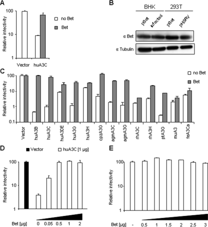
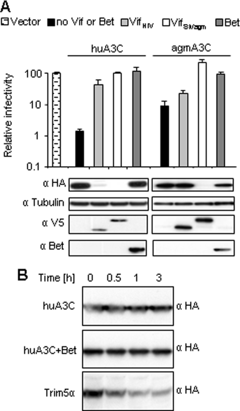
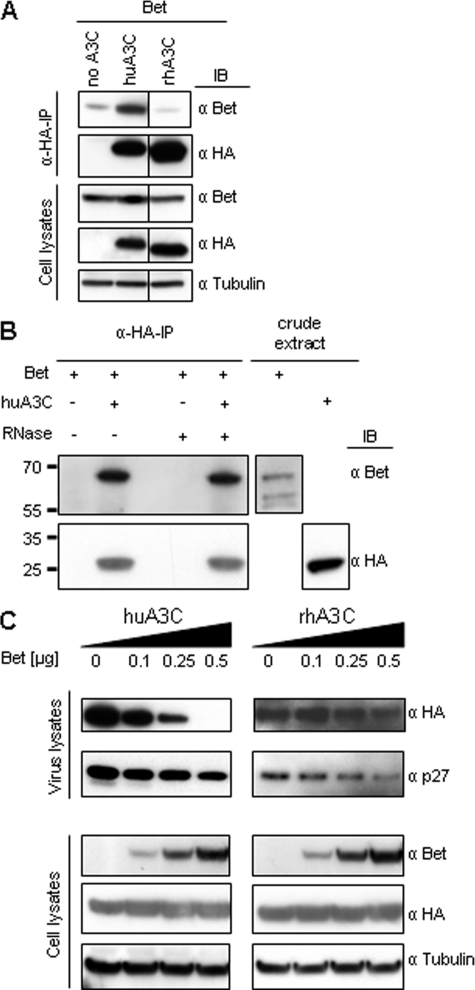
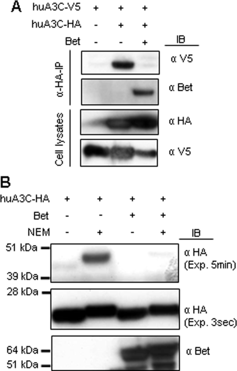
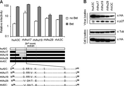
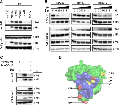
The APOBEC3 cytidine deaminases are part of the intrinsic defense of cells against retroviruses. Lentiviruses and spumaviruses have evolved essential accessory proteins, Vif and Bet, respectively, which counteract the APOBEC3 proteins. We show here that Bet of the Prototype foamy virus inhibits the antiviral APOBEC3C activity by a mechanism distinct to Vif: Bet forms a complex with APOBEC3C without inducing its degradation. Bet abolished APOBEC3C dimerization as shown by coimmunoprecipitation and cross-linking experiments. These findings implicate a physical interaction between Bet and the APOBEC3C. Subsequently, we identified the Bet interaction domain in human APOBEC3C in the predicted APOBEC3C dimerization site. Taken together, these data support the hypothesis that Bet inhibits incorporation of APOBEC3Cs into retroviral particles. Bet likely achieves this by trapping APOBEC3C protein in complexes rendering them unavailable for newly generated viruses due to direct immobilization.


Figures

FIGURE 1.
FIGURE 1.
Broad-range protection of SIV-based vectors by Bet to A3-mediated restriction. A, Bet-deficient lacZ-reporter viruses derived from PFV were produced in the absence (Vector) or presence of huA3C and Bet. Infectivity of normalized amounts of viruses was determined by quantification of β-galactosidase expression 3 days post infection (dpi). Infectivity, relative to the virus generated in absence of A3 and Bet, is given. B, the expression level of Bet in baby hamster kidney (BHK) cells was compared after transfection with Bet expression plasmid pBet and after infection with PFV at multiplicity of infection of 0.5 as well as in 293T cells after transfection with pBet and pHSRV (plasmid encoding full-length PFV genome). Cell lysis and immunoblot analysis were performed 2 days after transfection and infection, respectively. Bet was detected by using αBet antiserum. Equal loading of cell lysate samples was confirmed with α-tubulin antibody. C, Vif-deficient luciferase reporter viruses derived from SIVagm were produced in the absence (Vector) or presence of the indicated A3 and Bet proteins. Infectivity of equal amount of viruses was determined by quantification of luciferase activity 3 dpi. Infectivity, relative to the virus generated in absence of A3 and Bet, is shown. D, Δvif SIVagm-luc viruses were produced in the absence (Vector) or presence of huA3C and increasing amounts of Bet, or Bet alone (E). Infectivity of equal amount of viruses was determined by quantification of luciferase activity 3 dpi.
FIGURE 2.
FIGURE 2.
Bet inhibits A3C antiviral activity without leading to A3C degradation. A, Δvif SIVagm-luc viruses were produced in the presence or absence (Vector) of the indicated HA-tagged A3, V5-tagged VifHIV-1, and VifSIVagm and Bet expression vectors. Immunoblot analysis was performed with lysates of virus-producing cells. Expression of A3 proteins was detected with α-HA mAb, tubulin with α-tubulin mAb, Vif with α-V5 mAb, and Bet with Bet antiserum. Relative infectivity (relative to the virus generated in absence of A3 and Bet) of equal amounts of the reporter viruses is shown, demonstrating a close correlation of Vif-mediated degradation of A3 and protection, whereas Bet-mediated rescue is not associated with decreased A3 levels. Infectivity of viruses was determined by quantification of luciferase activity 3 dpi. B, 293T cells transiently expressing HA-tagged huA3C alone or together with Bet, and HA-tagged huTRIM5α were treated with cycloheximide. At the indicated time points after cycloheximide-induced suppression of protein biosynthesis, the cells were lysed and analyzed by immunoblotting using α-HA mAb.
FIGURE 3.
FIGURE 3.
Bet binds to huA3C and prevents its incorporation in virus particles. A, cells were transfected with Bet alone or with expression plasmids for HA-tagged huA3C and rhA3C. Cell lysates were prepared and α-HA-immunoprecipitation (α-HA-IP) was performed. A3-associated Bet was detected by immunoblot (IB) analysis using Bet antiserum. A3 proteins were detected in α-HA-IP samples and in cell lysates using α-HA mAbs, as well as Bet antiserum and α-tubulin antibodies in cell lysates. B, Bet-huA3C interaction occurs directly in an RNA-independent manner: Bet and huA3C were expressed in vitro using E. coli extracts, and α-HA-immunoprecipitations (α-HA-IP) were performed in the presence and absence of RNase A. Bet- and HA-tagged huA3C were detected in crude extracts and in immunoprecipitates by immunoblot analysis using Bet antiserum and α-HA mAb, respectively. C, virions were generated by co-transfection of Δvif SIVagm-luc, HA-tagged huA3C, or rhA3C and increasing amounts of Bet expression vector. Virions and cells were analyzed by immunoblot. A3C was detected with α-HA mAb, Bet with Bet antiserum, tubulin with α-tubulin mAb, and Capsid p27 protein with mAb to p24/p27.
FIGURE 4.
FIGURE 4.
Bet inhibits dimerization of huA3C. A, cells were transfected with V5-tagged huA3C alone or additionally with HA-tagged huA3C in presence or absence of Bet. Cell lysates were prepared, and α-HA-immunoprecipitation (α-HA-IP) was performed. huA3C-V5 co-precipitated with HA-tagged A3; Bet was detected by immunoblot (IB) analysis using Bet antiserum, huA3C-V5 using α-V5 mAb, and huA3C-HA using α-HA mAbs. B, lysates of cells expressing HA-tagged huA3C in the presence or absence of Bet were prepared and cross-linked with NEM. Monomeric and dimeric forms of huA3C were detected with α-HA mAb, whereas Bet was detected using Bet antiserum. Exp: exposure time.
FIGURE 5.
FIGURE 5.
Mapping of the Bet binding site within huA3C. A, Δvif SIVagmluc viruses were produced in the presence or absence of the indicated wt and rh/hu chimeric A3 and Bet proteins. Infectivity of equal amount of viruses was determined by quantification of luciferase activity, and is shown relative to virus generated in absence of A3 and Bet. Alignment of huA3C, rhA3C, and chimeric protein sequences (rh/hu17, -19, and -29) between aa 29 and 98 relative to the linear protein sequence with indicated Zn2+ coordination domain are shown. Identical aa to human and rhesus A3C are represented by a dashed line, aa that differ are shown, and human A3C-derived aa are highlighted in bold font. B, chimeric proteins are expressed and packaged into virions at similar level as parental huA3C and rhA3C. Expression and packaging were tested by immunoblot analysis of virus and cell lysates by using α-HA mAbs. Equal loading of samples was confirmed by detecting tubulin with α-tubulin mAb and Capsid p27 with mAb to p24/p27.
FIGURE 6.
FIGURE 6.
Bet interacts within the predicted dimerization site of huA3C. A, cells were transfected with Bet alone or with expression plasmids for HA-tagged huA3C, rhA3C, and indicated chimeras. Cell lysates were prepared, and α-HA-immunoprecipitation (α-HA-IP) was performed. A3-associated Bet was detected by immunoblot (IB) analysis using Bet antiserum. A3 proteins were detected in α-HA-IP samples and in cell lysates using α-HA mAbs, filters of cell lysates were also stained for Bet and tubulin. B, V5-tagged and HA-tagged huA3C (left panel), rhA3C (middle panel), and chimera rh/hu19 (right panel) were expressed in 293T cells in the presence of increasing amounts of Bet. Cell lysates were prepared, and α-HA-immunoprecipitation (α-HA-IP) was performed. Bet was detected by immunoblot (IB) analysis using Bet antiserum, V5-tagged proteins were detected using α-V5 mAb and HA-tagged proteins using α-HA mAbs. C, V5-tagged rh/hu19 was co-expressed with HA-tagged huA3C in the presence and absence of Bet. D, surface of huA3C modeled on the crystal structure of a complex of dimeric APOBEC2, including the predicted dimerization interface (blue), cysteine residues (yellow), and residues believed to mediate direct physical interaction with Bet (red).

Similar articles

Cited by

References

    1. Bieniasz, P. D. (2004) Nat. Immunol. 5 1109-1115 - PubMed
    1. Sawyer, S. L., Emerman, M., and Malik, H. S. (2004) PLoS Biol. 2 E275. - PMC - PubMed
    1. Münk, C., Beck, T., Zielonka, J., Hotz-Wagenblatt, A., Chareza, S., Battenberg, M., Thielebein, J., Cichutek, K., Bravo, I. G., O'Brien, S., Loechelt, M., and Yuhki, N. (2008) Genome Biology 9 R48. - PMC - PubMed
    1. Mbisa, J. L., Barr, R., Thomas, J. A., Vandegraaff, N., Dorweiler, I. J., Svarovskaia, E. S., Brown, W. L., Mansky, L. M., Gorelick, R. J., Harris, R. S., Engelman, A., and Pathak, V. K. (2007) J. Virol. 81 7099-7110 - PMC - PubMed
    1. Luo, K., Wang, T., Liu, B., Tian, C., Xiao, Z., Kappes, J., and Yu, X. F. (2007) J. Virol. 81 7238-7248 - PMC - PubMed

Publication types

MeSH terms

Associated data

LinkOut - more resources