. 2009 Feb 20;284(8):4829-35.
doi: 10.1074/jbc.M807943200. Epub 2008 Dec 12.

Redox control of the DNA damage-inducible protein DinG helicase activity via its iron-sulfur cluster

Affiliations

Redox control of the DNA damage-inducible protein DinG helicase activity via its iron-sulfur cluster

Binbin Ren et al. J Biol Chem. .

Abstract

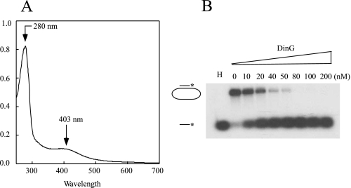
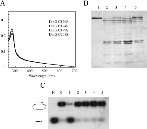
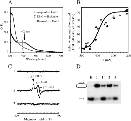
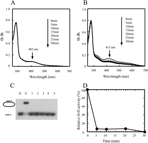
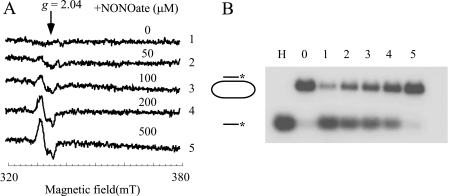
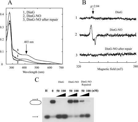
The Escherichia coli DNA damage-inducible protein DinG, a member of the superfamily 2 DNA helicases, has been implicated in the nucleotide excision repair and recombinational DNA repair pathways. Combining UV-visible absorption, EPR, and enzyme activity measurements, we demonstrate here that E. coli DinG contains a redox-active [4Fe-4S] cluster with a midpoint redox potential (E(m)) of -390 +/- 23 mV (pH 8.0) and that reduction of the [4Fe-4S] cluster reversibly switches off the DinG helicase activity. Unlike the [4Fe-4S] cluster in E. coli dihydroxyacid dehydratase, the DinG [4Fe-4S] cluster is stable, and the enzyme remains fully active after exposure to 100-fold excess of hydrogen peroxide, indicating that DinG could be functional under oxidative stress conditions. However, the DinG [4Fe-4S] cluster can be efficiently modified by nitric oxide (NO), forming the DinG-bound dinitrosyl iron complex with the concomitant inactivation of helicase activity in vitro and in vivo. Reassembly of the [4Fe-4S] cluster in NO-modified DinG restores helicase activity, indicating that the iron-sulfur cluster in DinG is the primary target of NO cytotoxicity. The results led us to propose that the iron-sulfur cluster in DinG may act as a sensor of intracellular redox potential to modulate its helicase activity and that modification of the iron-sulfur cluster in DinG and likely in other DNA repair enzymes by NO may contribute to NO-mediated genomic instability.


Figures

FIGURE 1.
FIGURE 1.
Purified E. coli DinG contains an iron-sulfur cluster. A, UV-visible absorption spectrum of purified E. coli DinG. The protein concentration was ∼10 μm. B, the helicase activity of purified E. coli DinG. Purified DinG (at a final concentration of 0–200 nm) was incubated with the 32P-radioactively labeled substrate in the presence of ATP (2 mm) at 30 °C for 10 min. The reaction product (single-stranded DNA) was separated by agarose gel (1%) electrophoresis as described under “Experimental Procedures.” In lane H, the sample was heated at 85 °C for 5 min. The concentration of DinG in the reaction solution is indicated at the top of each lane.
FIGURE 2.
FIGURE 2.
The conserved cysteine residues in DinG are required for iron-sulfur cluster binding, protein stability, and helicase activity. The DinG mutants (C120S, C194S, C199S, and C205S) were constructed and purified as described under “Experimental Procedures.” A, UV-visible absorption spectra of purified DinG mutants. The protein concentrations were ∼3 μm. B, SDS-PAGE analysis of purified wild-type DinG and DinG mutants. The same amount of cells containing either wild-type DinG or the DinG mutants was used for protein purification. Equal amounts of purified proteins were analyzed on the SDS-polyacrylamide gel. Lanes 1–5, wild-type DinG and mutants C120S, C194S, C199S, and C205S, respectively. C, the helicase activity of purified wild-type DinG and the DinG mutants. Purified proteins (200 nm) were incubated with the 32P-radioactively labeled substrate in the presence of ATP (2 mm) at 30 °C for 10 min. The reaction product (single-stranded DNA) was separated by agarose gel (1%) electrophoresis as described under “Experimental Procedures.” Lane H, sample heated at 85 °C for 5 min; lane 0, no enzyme added; lanes 1–5, wild-type DinG and mutants C120S, C194S, C199S, and C205S, respectively.
FIGURE 3.
FIGURE 3.
Redox state of the iron-sulfur cluster in DinG regulates its helicase activity. A, UV-visible absorption spectra of reduced and oxidized DinG. Purified DinG (25 μm; spectrum 1) was reduced with sodium dithionite (200 μm) under anaerobic conditions (spectrum 2) and then reoxidized by exposure to air for 30 min (spectrum 3). The absorption peak at 403 nm reflects the oxidized DinG [4Fe-4S] cluster. B, redox titration of the DinG [4Fe-4S] cluster. Purified DinG (25 μm) in buffer containing Tris (50 mm, pH 8.0) and NaCl (500 mm) was supplemented with safranin O (1 μm) as a redox mediator. Redox titration was performed in an anaerobic redox cuvette as described under “Experimental Procedures.” The x axis shows the redox potentials measured with a redox microelectrode. The y axis shows the relative absorbance at 403 nm, normalized to 0 or 100% for a fully reduced or oxidized DinG [4Fe-4S] cluster, respectively. The solid line drawn through the data points represents the best fit to the Nernst equation (n = 1) with Em of –390 ± 23 mV. Data were from three independent experiments represented with three different symbols. C, EPR spectra of purified DinG. Purified DinG (500 μm) (spectrum 1) was reduced with sodium dithionite (2 mm; spectrum 2) or reoxidized with potassium ferricyanide (2 mm; spectrum 3). D, the helicase activity of DinG under different redox potentials. Purified DinG (100 nm) was either reduced with sodium dithionite or reoxidized with potassium ferricyanide before the 32P-radioactively labeled DNA substrate was added to the incubation solutions. After a 5-min incubation at 30 °C, the reaction was terminated, and the product (single-stranded DNA) was separated by agarose gel (1%) electrophoresis as described under “Experimental Procedures.” Lane H, sample heated at 85 °C for 5 min; lane 0, no enzyme added; lane 1, purified DinG; lane 2, purified DinG reduced with dithionite (1.0 mm); lane 3, dithionite-reduced DinG reoxidized with potassium ferricyanide (2 mm).
FIGURE 4.
FIGURE 4.
The iron-sulfur cluster in DinG is resistant to hydrogen peroxide. A, effect of H2O2 on the DinG [4Fe-4S] cluster. Purified DinG (10 μm) was incubated with H2O2 (1 mm) at 25 °C. UV-visible spectra were taken every 5 min after addition of H2O2 for 30 min. B, effect of H2O2 on the dihydroxyacid dehydratase [4Fe-4S] cluster. Purified E. coli dihydroxyacid dehydratase (20 μm) was incubated with hydrogen peroxide (1 mm) at 25 °C. UV-visible spectra were taken every 5 min after addition of H2O2 for 30 min. C, effect of H2O2 on the DinG helicase activity. After incubation with H2O2 for the indicated time, DinG (at a final concentration of 100 nm) was used for the helicase activity assay. Lane H, sample heated at 85 °C for 5 min; lane 0, no enzyme added; lanes 1–5, purified DinG after incubation with H2O2 for 0, 5, 10, 20, and 30 min, respectively. The reaction product (single-stranded DNA) was separated by agarose gel (1%) electrophoresis as described under “Experimental Procedures.” D, effect of H2O2 on the enzyme activity of dihydroxyacid dehydratase. The relative enzyme activity of dihydroxyacid dehydratase after incubation with H2O2 (1 mm) was measured as described under “Experimental Procedures” and plotted as a function of incubation time with H2O2.
FIGURE 5.
FIGURE 5.
The DinG iron-sulfur cluster is sensitive to NO. A, modification of the DinG [4Fe-4S] cluster by NO. Purified DinG (30 μm) was incubated with different amounts of diethylamine NONOate in buffer containing Tris (20 mm, pH 7.5) and NaCl (200 mm) at room temperature under anaerobic conditions. After a 20-min incubation, protein was repurified by passage through a HiTrap desalting column. Spectra 1–5, purified DinG incubated with 0, 50, 100, 200, and 500 μm NONOate under anaerobic conditions. The protein concentrations of repurified DinG were ∼4 μm. mT, milliteslas. B, inactivation of DinG helicase activity by NO. After incubation with different amounts of NONOate, repurified DinG (at a final concentration of 100 nm) was used for the helicase activity assay. Lane H, sample heated at 85 °C for 5 min; lane 0, no enzyme added; lanes 1–5, repurified DinG after incubation with 0, 50, 100, 200, and 500 μm NONOate. The reaction product (single-stranded DNA) was separated by agarose gel (1%) electrophoresis as described under “Experimental Procedures.”
FIGURE 6.
FIGURE 6.
Reactivation of NO-modified DinG by reassembly of iron-sulfur clusters. Purified DinG (30 μm) was exposed to NO (0.5 mm NONOate) under anaerobic conditions, followed by repair using the iron-sulfur cluster repair system as described under “Experimental Procedures.” A, UV-visible absorption spectra of DinG. Spectrum 1, purified DinG before NO exposure (DinG); spectrum 2, purified DinG after NO exposure (DinG-NO); spectrum 3, NO-exposed DinG repaired with the iron-sulfur cluster repair system (DinG-NO repaired). The protein concentrations of DinG were ∼5 μm. B, EPR spectra of DinG. Spectrum 1, purified DinG before NO exposure; spectrum 2, purified DinG after NO exposure; spectrum 3, NO-exposed DinG repaired with the iron-sulfur cluster assembly system. The protein concentrations of DinG were ∼5 μm. mT, milliteslas. C, reversible inactivation of DinG by NO. Two concentrations of DinG (50 and 100 nm) were used for the helicase activity assay. In lane H, the sample was heated at 85 °C for 5 min. In lane 0, no enzyme was added. The reaction product (single-stranded DNA) was separated by agarose gel (1%) electrophoresis as described under “Experimental Procedures.”

Similar articles

Cited by

References

    1. Lewis, L. K., Jenkins, M. E., and Mount, D. W. (1992) J. Bacteriol. 174 3377–3385 - PMC - PubMed
    1. Van Dyk, T. K., DeRose, E. J., and Gonye, G. E. (2001) J. Bacteriol. 183 5496–5505 - PMC - PubMed
    1. Fernandez De Henestrosa, A. R., Ogi, T., Aoyagi, S., Chafin, D., Hayes, J. J., Ohmori, H., and Woodgate, R. (2000) Mol. Microbiol. 35 1560–1572 - PubMed
    1. Koonin, E. V. (1993) Nucleic Acids Res. 21 1497. - PMC - PubMed
    1. Voloshin, O. N., Vanevski, F., Khil, P. P., and Camerini-Otero, R. D. (2003) J. Biol. Chem. 278 28284–28293 - PubMed

Publication types

MeSH terms