. 2009 Feb 13;284(7):4267-74.
doi: 10.1074/jbc.M807665200. Epub 2008 Dec 11.

A role for the proton-coupled folate transporter (PCFT-SLC46A1) in folate receptor-mediated endocytosis

Affiliations

A role for the proton-coupled folate transporter (PCFT-SLC46A1) in folate receptor-mediated endocytosis

Rongbao Zhao et al. J Biol Chem. .

Abstract

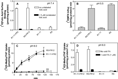
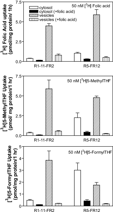
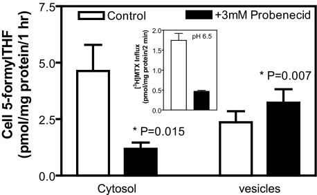
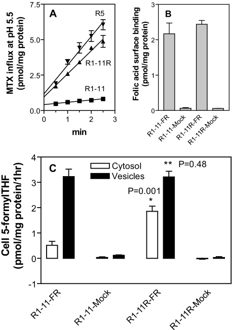
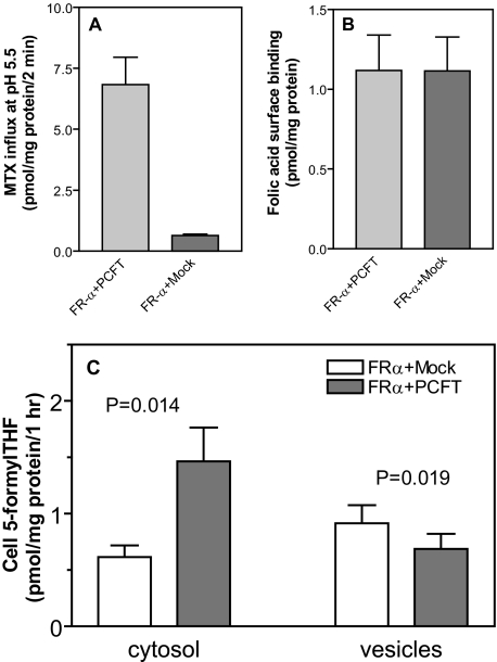
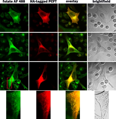
Recently, this laboratory identified a proton-coupled folate transporter (PCFT), with optimal activity at low pH. PCFT is critical to intestinal folate absorption and transport into the central nervous system because there are loss-of-function mutations in this gene in the autosomal recessive disorder, hereditary folate malabsorption. The current study addresses the role PCFT might play in another transport pathway, folate receptor (FR)-mediated endocytosis. FRalpha cDNA was transfected into novel PCFT(+) and PCFT(-) HeLa sublines. FRalpha was shown to bind and trap folates in vesicles but with minimal export into the cytosol in PCFT(-) cells. Cotransfection of FRalpha and PCFT resulted in enhanced folate transport into cytosol as compared with transfection of FRalpha alone. Probenecid did not inhibit folate binding to FR, but inhibited PCFT-mediated transport at endosomal pH, and blocked FRalpha-mediated transport into the cytosol. FRalpha and PCFT co-localized to the endosomal compartment. These observations (i) indicate that PCFT plays a role in FRalpha-mediated endocytosis by serving as a route of export of folates from acidified endosomes and (ii) provide a functional role for PCFT in tissues in which it is expressed, such as the choroid plexus, where the extracellular milieu is at neutral pH.


Figures

FIGURE 1.
FIGURE 1.
Comparison of folate transport in HeLa R1-11 (PCFT) and HeLa R5 (PCFT+) cells in the presence and absence of FRα expression. Panel A, FRα expression as determined by the difference in [3H]folic acid (5 nm) surface binding at pH 7.4 in the presence and absence of 5 μm (1000-fold in excess) nonlabeled folic acid. Panel B, level of PCFT-functional expression as assessed by [3H]MTX (0.5 μm) influx directly across the plasma membrane over 2 min at pH 5.5. Panel C, FRα-mediated uptake of 5-[3H]methylTHF (50 nm) at pH 8.0. Under these conditions transport directly across the plasma membrane mediated by PCFT is minimized; uptake occurs almost entirely by receptor-mediated endocytosis. Panel D, specificity of 5-methylTHF transport mediated by FRα at pH 8.0 was verified by co-addition of 1 μm nonlabeled folic acid that blocked FRα-mediated endocytosis. Data for all panels are the mean ± S.E. from three independent experiments.
FIGURE 2.
FIGURE 2.
Discrimination of tritiated folates in cytosol and vesicles following transport mediated by FRα. Cells were exposed to 50 nm folic acid (upper panel), 5-methylTHF (middle panel), or 5-formylTHF (lower panel) for 1 h at pH 8.0. After lysis of cells in hypotonic buffer, the vesicle and cytosolic fractions were separated by centrifugation. The symbols for the figures, indicated in the upper panel, is the same for all panels. Co-addition of 1 μm folic acid was utilized to suppress FRα-mediated transport. Data for all panels are the mean ± S.E. from three independent experiments.
FIGURE 3.
FIGURE 3.
Effect of 3 mm probenecid on partition of transported 5-formylTHF between cytosol and vesicles in R5-FR12 cells. Cells were exposed to 50 nm 5-[3H]formylTHF for 1 h at pH8.0 in the presence or absence of 3 mm probenecid. The data represent transport mediated specifically by FRα; the component not inhibited by 1 μm nonlabeled folic acid was subtracted, as indicated in Fig. 2. The inset illustrates the effect of 3 mm probenecid on PCFT-mediated transport across the plasma membrane. This was assessed with 0.5 μm [3H]MTX at pH 6.5 over 2 min to simulate the impact of probenecid on PCFT-mediated export from endosomes. Data are the mean ± S.E. from five independent experiments.
FIGURE 4.
FIGURE 4.
Transport in R1-11 and the PCFT-reverted R1-11R cells transiently transfected with FRα. Panel A, PCFT functional expression was assessed in R1-11, R1-11R, and R5 cells by measurement of [3H]MTX (0.5 μm) influx at pH 5.5. Panel B, FRα levels assayed by [3H]folic acid surface binding in cells transiently transfected with FRα cDNA or vector only (Mock). Panel C, FRα-mediated 5-[3H]formylTHF transport into vesicles and cytosol in transient transfectants. *, Student's t test between cytosol values. **, Student's t test between vesicle values. Data for all panels are the mean ± S.E. from three independent experiments.
FIGURE 5.
FIGURE 5.
Comparison of FRα-mediated transport in R1-11 cells transiently transfected with FRα cDNA, alone, or co-transfected with PCFT cDNA. Panel A, PCFT functional expression as assessed by [3H]MTX (0.5 μm) influx at pH 5.5. Panel B, FRα expression as assessed by [3H]folic acid surface binding. Panel C, FRα-mediated 5-[3H]formylTHF transport into vesicles and cytosol. Data for all panels are the mean ± S.E. from four independent experiments.
FIGURE 6.
FIGURE 6.
Localization of endogenous PCFT in murine choroid plexus. PCFT was visualized with a polyclonal antibody to a peptide derived from the C terminus of the murine protein. Slides were counterstained with Mayer's hematoxylin. The immunohistochemical procedures are described in detail under “Experimental Procedures.”
FIGURE 7.
FIGURE 7.
Subcellular co-localization of folate-AlexaFluor 488 and PCFT. R1-11-FR2 cells stably transfected with human PCFT were incubated with 100 nm folate-AlexaFluor 488 for 60 min at 37 °C. After washing to remove unbound folate-AlexaFluor 488, cells were incubated in fresh medium for 1 h before fixation and examination by confocal microscopy (see under “Experimental Procedures”). Three representative cells (rows 1–3) are shown. Column 1 indicates the localization of the internalized folate-AlexaFluor 488 (green). Column 2 indicates the localization of HA-tagged endogenous PCFT visualized with an anti-HA antibody (red). Co-localization of the two markers is seen in the overlay (yellow) in column 3. A magnification of a portion of each cell (marked by the red box in row 3) is indicated in row 4.

Similar articles

Cited by

References

    1. Qiu, A., Jansen, M., Sakaris, A., Min, S. H., Chattopadhyay, S., Tsai, E., Sandoval, C., Zhao, R., Akabas, M. H., and Goldman, I. D. (2006) Cell 127 917–928 - PubMed
    1. Zhao, R., Min, S. H., Qiu, A., Sakaris, A., Goldberg, G. L., Sandoval, C., Malatack, J. J., Rosenblatt, D. S., and Goldman, I. D. (2007) Blood 110 1147–1152 - PMC - PubMed
    1. Min, S. H., OH, S. Y., Karp, G. I., Poncz, M., Zhao, R., and Goldman, I. D. (2008) J. Pediatr. 153 435–437 - PMC - PubMed
    1. Lasry, I., Berman, B., Straussberg, R., Sofer, Y., Bessler, H., Sharkia, M., Glaser, F., Jansen, G., Drori, S., and Assaraf, Y. G. (2008) Blood 112 2055–2061 - PubMed
    1. Geller, J., Kronn, D., Jayabose, S., and Sandoval, C. (2002) Medicine (Baltimore) 81 51–68 - PubMed

Publication types

MeSH terms