. 2009 Feb;106(2):620-30.
doi: 10.1152/japplphysiol.91277.2008. Epub 2008 Dec 12.

Long-loop pathways in cardiovascular electroacupuncture responses

Affiliations

Long-loop pathways in cardiovascular electroacupuncture responses

Peng Li et al. J Appl Physiol (1985). 2009 Feb.

Abstract

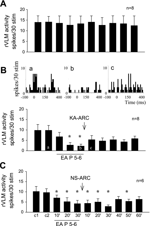
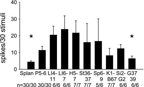
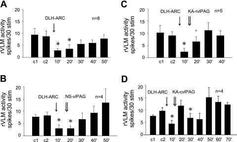
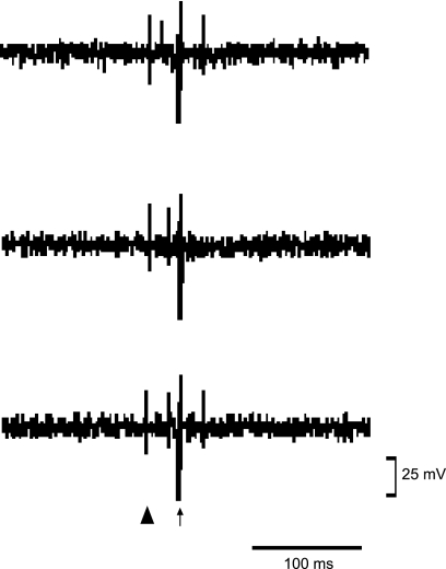
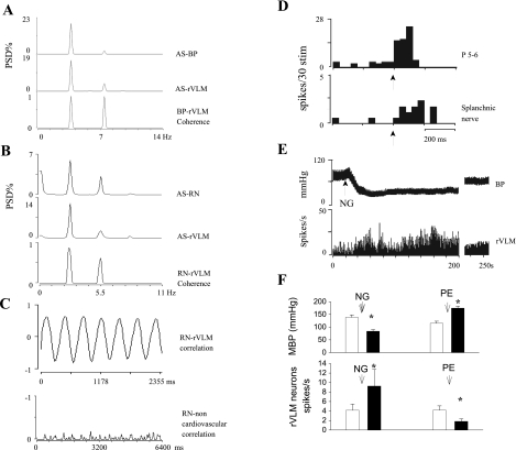
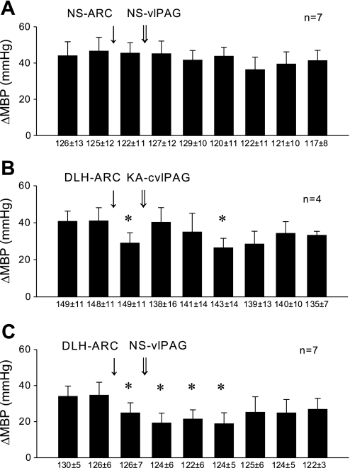
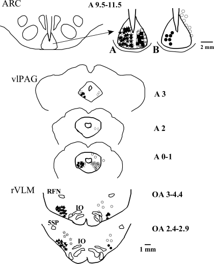
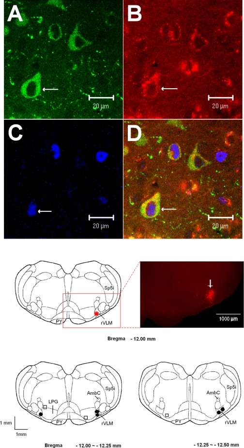
We have shown that electroacupuncture (EA) at P 5-6 (overlying median nerves) activates arcuate (ARC) neurons, which excite the ventrolateral periaqueductal gray (vlPAG) and inhibit cardiovascular sympathoexcitatory neurons in the rostral ventrolateral medulla (rVLM). To investigate whether the ARC inhibits rVLM activity directly or indirectly, we stimulated the splanchnic nerve to activate rVLM neurons. Micropipettes were inserted in the rVLM, vlPAG, and ARC for neural recording or injection. Microinjection of kainic acid (KA; 1 mM, 50 nl) in the ARC blocked EA inhibition of the splanchnic nerve stimulation-induced reflex increases in rVLM neuronal activity. Microinjection of d,l-homocysteic acid (4 nM, 50 nl) in the ARC, like EA, inhibited reflex increases in the rVLM neuronal discharge. The vlPAG neurons receive convergent input from the ARC, splanchnic nerve, P 5-6, and other acupoints. Microinjection of KA bilaterally into the rostral vlPAG partially reversed rVLM neuronal responses and cardiovascular inhibition during d,l-homocysteic acid stimulation of the ARC. On the other hand, injection of KA into the caudal vlPAG completely reversed these responses. We also observed that ARC neurons could be antidromically activated by stimulating the rVLM, and that ARC perikarya was labeled with retrograde tracer that had been microinjected into the rVLM. These neurons frequently contained beta-endorphin and c-Fos, activated by EA stimulation. Therefore, the vlPAG, particularly, the caudal vlPAG, is required for ARC inhibition of rVLM neuronal activation and subsequent EA-related cardiovascular activation. Direct projections from the ARC to the rVLM, which serve as an important source of beta-endorphin, appear also to exist.


Figures

Fig. 1.
Fig. 1.
Bilateral microinjection of kainic acid (KA) in arcuate (ARC) nucleus blocked electroacupuncture (EA) inhibition of rostral ventrolateral medulla (rVLM) neuronal response to splanchnic nerve stimulation. A: time control of rVLM response to stimulation of splanchnic nerve every 10 min. B, bottom: bilateral microinjection of KA into the ARC nucleus rapidly reversed EA inhibition of the rVLM neural response. *P < 0.05 compared with controls. Top: peristimulus time histograms demonstrate ventrolateral periaqueductal gray (vlPAG) neuronal responses during repeated 15-s (2-Hz) splanchnic nerve stimulations before (a) and after EA (b) at P 5–6 and following KA injection into the ARC (c). C: EA applied bilaterally at P 5–6 acupoints (located over median nerve) for 30 min inhibited splanchnic nerve stimulation-induced rVLM neural responses for more than 50 min. Microinjection of normal saline (NS) into the ARC did not influence the EA-related inhibition.
Fig. 2.
Fig. 2.
Convergent activity in the vlPAG between A 0.1–3. Bar histograms show evoked activity in vlPAG neurons during individual stimulation of the splanchnic nerve and nine sets of acupoints. Numbers below each bar indicate the number of responsive cells relative to number of cells studied. *Significantly different discharge rate compared with response during stimulation of P 5–6 (P < 0.05).
Fig. 3.
Fig. 3.
Histograms displaying the effect of bilateral microinjection of d,l-homocysteic acid (DLH) and KA in ARC on the rVLM-evoked response to splanchnic nerve stimulation. A: microinjection of DLH in the ARC inhibited rVLM response for 20 min. B: microinjection of NS (vehicle) in the rostral (rvlPAG; n = 2) or caudal (cvlPAG; n = 2) vlPAG did not influence the inhibitory effect resulting from stimulation of the ARC with DLH. C: microinjection of KA in the rvlPAG partially reversed the inhibitory rVLM effect caused by microinjection of DLH in the ARC. D: microinjection of KA in the cvlPAG fully reversed the inhibitory rVLM effect of microinjection of DLH in the ARC *P < 0.05 compared with controls (c1, c2).
Fig. 4.
Fig. 4.
Tracing showing a single-unit recording of neural activity in ARC evaluating the response to antidromic stimulation of rVLM. Top: first spike is the orthodromic evoked potential induced during stimulation of P 5–6; second spike represents the response to antidromic stimulation of rVLM. Middle: the P 5–6-induced spike moved toward the rVLM antidromic stimulation and canceled the antidromic response. Bottom: the spike induced by stimulation of P 5–6 moved backward, and the antidromic response reappeared. ▴ and ↑ represent the stimulation artifacts of P 5–6 and rVLM, respectively.
Fig. 5.
Fig. 5.
Illustrations showing identification of cardiovascular sympathetic neurons in rVLM. A: autospectra (AS) of blood pressure (BP) and rVLM neuronal activity and the corresponding coherence function. Coherence was 0.87 at 3.79 Hz. B: AS of renal nerve (RN) and rVLM neuronal activity and the corresponding coherence of RN and rVLM neuronal firings. Coherence of 0.86 occurred at a frequency of 3.3 Hz. C, top: cross correlation of RN discharge and rVLM neural activity. Bottom: a neuron outside rVLM, showing no correlation with RN activity. D: histograms show the rVLM neuronal responses to stimulation of P 5–6 and splanchnic nerve. ↑, Stimulation of P 5–6 or splanchnic nerve. E: the change in BP and spontaneous discharge of an rVLM neuron after intravenous administration of nitroglycerin (NG). F: histograms show the change of BP and rVLM neuronal discharge after intravenous injection of NG and phenylephrine (PE) in 15 cats. MBP, mean BP; PSD, power spectral density. *, <0.05 compared with controls.
Fig. 6.
Fig. 6.
Histograms illustrating hemodynamic responses. A: repeated reflex BP responses were induced by application of bradykinin (BK) on gallbladder every 10 min. The responses were not altered by microinjection of NS into the rvlPAG or cvlPAG. B: inhibition of reflex BP response by bilateral injections of DLH in the ARC nucleus was blocked by bilateral injection of KA in the cvlPAG. C: inhibition of reflex BP response by injections of DLH in the ARC nucleus was not altered by bilateral injection of NS in the rvlPAG or cvlPAG. Numbers below the baseline indicate resting MBP, which were unchanged by the protocols. Δ, Change. *P < 0.05 compared with controls.
Fig. 7.
Fig. 7.
Composite map of brain sections showing locations of microinjection, stimulation, and recording sites in ARC, vlPAG, and rVLM. For simplicity, all recordings or microinjections are placed on one side. Top, left: location of the ARC in the ventral hypothalamus. Right: A: ★, DLH in ARC, n = 30; ▪, KA in ARC, n = 6; □, NS in ARC, n = 13. B: •, antidromic recordings in ARC, n = 6; ○, hypothalamic neurons unresponsive to antidromic stimulation of rVLM, n = 8. Middle: ▪, KA in rvlPAG (n = 5) and in cvlPAG (n = 8); □, NS in vlPAG, n = 16; ◊, recording of neuronal response to stimulation of the splanchnic nerve and somatic acupoints (n = 30); ○, recordings outside vlPAG at A 0.1–3 level (n = 3, another unresponsive neuron located more rostrally, A 7) is not shown in this figure. Bottom: ▴, rVLM neurons responding to EA, n = 12; ▾, rVLM evaluated for time control, n = 8; ★, rVLM neurons inhibited by DLH in ARC, n = 12; ⧫, DLH in ARC, KA in rvlPAG (n = 5) and cvlPAG (n = 4), and recording in rVLM; ⋆, medullary neurons outside rVLM unresponsive to DLH in ARC; n = 5; •, antidromic stimulation in rVLM, n = 6; ○, antidromic stimulation outside rVLM, n = 8. Retrofacial nucleus (RFN), nucleus of the inferior olive (IO), and alaminar spinal trigeminal nucleus (5SP) are indicated.
Fig. 8.
Fig. 8.
AD: confocal microscopic images displaying β-endorphin, retrogradely transported microsphere tracer injected 10 days earlier into rVLM and c-Fos, in the ARC nucleus (Bregma −1.72 mm) from a rat treated with EA. AC: immunostaining of β-endorphin (green; A), retrograde microsphere tracer (red; B), and c-Fos (blue; C). D: merged images from AC. Arrows in AD indicate a neuron containing β-endorphin, microsphere c-Fos, and β-endorphin + microspheres + c-Fos, respectively. Middle: an original picture (right) represents the area indicated in the box on the left. This fluorescent microscopic image demonstrates the injection site of the retrograde microsphere tracer in the rVLM (bregma −12 mm) of a rat following EA. The bright red area indicated by the arrow shows an injected site of rhodamine-labeled fluorescent microsphere in the rVLM. Scale bar = 1,000 μm. Bottom: sections of rat medulla oblongata illustrating microinjection sites of the retrograde microsphere tracer. • and □ indicate injection sites inside and outside the rVLM, respectively. AmbC, ambig compact; LPG, lateral paragigantocellularis nucleus; PY, pyramidal tract; Sp5i, spinal 5 interpolar nucleus.

References

    1. Barman S, Gebber G. Sequence of activation of ventrolateral and dorsal medullary sympathetic neurons. Am J Physiol Regul Integr Comp Physiol 245: R438–R447, 1983. - PubMed
    1. Berman AL The Brainstem of the Cat: A Cytoarchitectonic Atlas With Stereotaxic Coordinates. Madison, WI: University of Wisconsin Press, 1968.
    1. Bloom F, Battenberg E, Rossier J, Ling N, Guillemin R. Neurons containing B-endorphin in rat brain exist separately from those containing enkephalin: immunocytochemical studies. Proc Natl Acad Sci USA 75: 1591–1595, 1978. - PMC - PubMed
    1. Bureš J, Petran M, Zachar J. Electrophysiological Methods in Biological Research. New York: Academic, 1967.
    1. Carrive P, Bandler R. Viscerotopic organization of neurons subserving hypotensive reactions within the midbrain periaqueductal: a correlative functional and anatomical study. Brain Res 541: 206–216, 1991. - PubMed

Publication types

MeSH terms

LinkOut - more resources