Review
. 2009 Jan;174(1):3-13.
doi: 10.2353/ajpath.2009.071151. Epub 2008 Dec 12.

Where asthma and hypersensitivity pneumonitis meet and differ: noneosinophilic severe asthma

Affiliations
Review

Where asthma and hypersensitivity pneumonitis meet and differ: noneosinophilic severe asthma

Pieter Bogaert et al. Am J Pathol. 2009 Jan.

Abstract

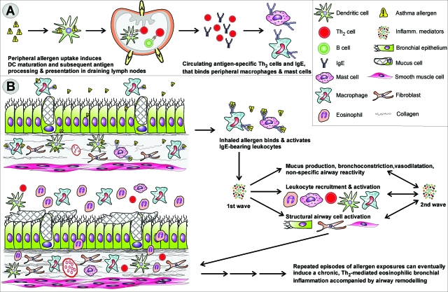
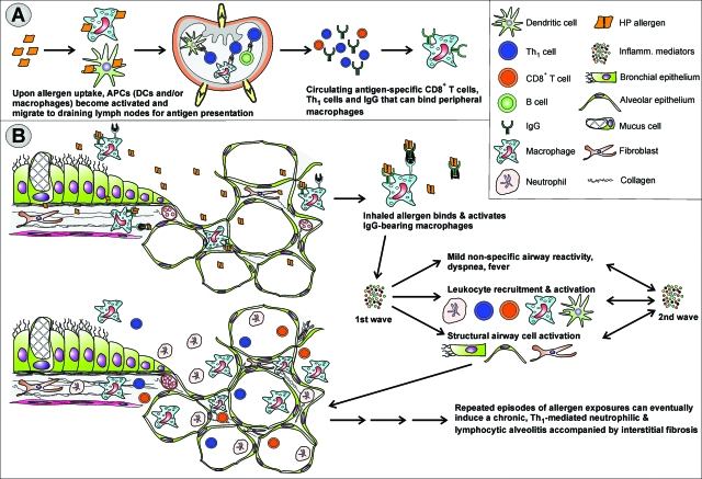
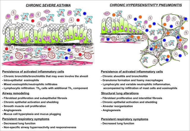
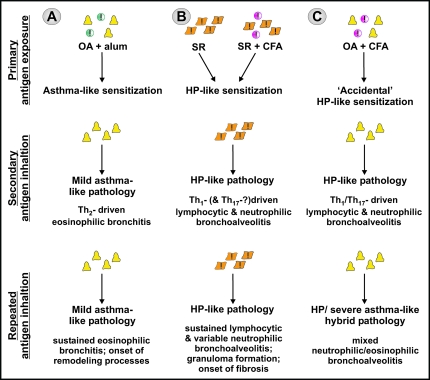
Asthma is a type-I allergic airway disease characterized by Th(2) cells and IgE. Episodes of bronchial inflammation, eosinophilic in nature and promoting bronchoconstriction, may become chronic and lead to persistent respiratory symptoms and irreversible structural airway changes. Representative mostly of mild to moderate asthma, this clinical definition fails to account for the atypical and often more severe phenotype found in a considerable proportion of asthmatics who have increased neutrophil cell counts in the airways as a distinguishing trait. Neutrophilic inflammation is a hallmark of another type of allergic airway pathology, hypersensitivity pneumonitis. Considered as an immune counterpart of asthma, hypersensitivity pneumonitis is a prototypical type-III allergic inflammatory reaction involving the alveoli and lung interstitium, steered by Th(1) cells and IgG and, in its chronic form, accompanied by fibrosis. Although pathologically very different and commonly approached as separate disorders, as discussed in this review, clinical studies as well as data from animal models reveal undeniable parallels between both airway diseases. Danger signaling elicited by the allergenic agent or by accompanying microbial patterns emerges as critical in enabling immune sensitization and in determining the type of sensitization and ensuing allergic disease. On this basis, we propose that asthma allergens cause severe noneosinophilic asthma because of sensitization in the presence of hypersensitivity pneumonitis-promoting danger signaling.


Figures

Figure 1
Figure 1
Development of mild asthma. A: On primary allergen exposure, DAMPs and/or PAMPs intrinsic to or accompanying the allergen activate DCs to become APCs biased toward the induction of Th2 cells. As a result, immune sensitization featuring allergen-specific IgE and Th2 cells is generated. B: Activation of IgE-bearing innate cells by inhaled allergen triggers the release of pro-inflammatory mediators such as histamine, leukotrienes, Th2-associated cytokines and chemokines, and reactive oxygen species. The resulting early-phase allergic reaction is manifested by smooth muscle contraction, mucus hypersecretion, and increased airway responsiveness. Inflammatory cell recruitment and activation leads to the production of a second wave of particularly Th2-associated inflammatory mediators such as IL-4, IL-5, IL-13, eotaxins, RANTES, and others. This late-phase reaction occurs ∼6 to 9 hours after allergen inhalation and usually lasts for not more than a day.
Figure 2
Figure 2
Development of HP. A: Primary allergen exposure and subsequent naïve CD4+ T-cell stimulation by activated APCs—alveolar macrophages and/or lung DCs—results in the generation of allergen-specific Th1 cells and IgG. Danger signals elicited by PAMPs intrinsic to the HP allergen and/or present at the time of exposure are crucial in generating the Th1-bias of sensitization. B: Airway inflammation is initiated on secondary allergen exposure in the alveoli. IgG-allergen immune complexes trigger the complement cascade and/or directly activate alveolar macrophages to produce reactive oxygen species and inflammatory cytokines such as tumor necrosis factor-α and IL-12. These in turn trigger the production of IFN-γ by Th1 cells, resulting in a highly inflammatory cytokine mix that along with various chemokines such as IL-8, IP-10, I-TAC, and MIG, is at the basis of the neutrophilic nature of the inflammatory cell infiltrate, characteristic of HP.
Figure 3
Figure 3
(Dis-)similar features of chronic severe asthma and chronic HP. Although the site, nature, and regulation of the inflammatory response in both diseases differ greatly, the chronic stages of severe asthma and HP show some remarkable similarities. In both conditions the chronic inflammatory state is induced by repeated episodes of allergen exposures and subsequent inflammatory responses. The resulting sustained production of pro-inflammatory mediators and growth factors generates a self-perpetuating process of inflammatory cell survival and accumulation, chronic epithelial activation, and deregulation of normal healing processes, accompanied by persistent respiratory symptoms and decrease in lung function. Moreover, the distinction between Th2-driven airway eosinophilia versus Th1-driven neutrophilic alveolitis becomes blurred: the inflammatory cell infiltrate contains a significant fraction of neutrophils in chronic severe asthma and eosinophils and mast cells in chronic HP.
Figure 4
Figure 4
Experimental mouse models illustrate the critical role of accompanying adjuvant activity in determining the type of immune sensitization and ensuing allergic disease. A: Systemic sensitization against inert OA in the presence of alum predisposes to a Th2-driven eosinophilic bronchoalveolitis. Repeated OA exposures in the absence of further immunogenic stimuli sustain the eosinophilic inflammatory airway response, much like mild asthma. B: Intranasal instillation of S. rectivergula or systemic immunization in the presence of CFA predisposes for a Th1- and possibly Th17-driven neutrophilic bronchoalveolitis. Like HP, repeated S. rectivergula inhalations further sustain this inflammatory response, presumably through the intrinsic adjuvant property of S. rectivergula. C: Systemic sensitization against OA in the presence of CFA similarly predisposes to a Th1- and Th17-driven neutrophilic bronchoalveolitis. On further exposures, the intrinsically inert nature of the antigen along with the absence of Th1-promoting stimuli supposedly is at the basis of the inflammatory pattern evolving to a mixed neutrophilic and eosinophilic inflammation, much like severe noneosinophilic asthma.

References

    1. Wills-Karp M, Santeliz J, Karp CL. The germless theory of allergic disease: revisiting the hygiene hypothesis. Nat Rev Immunol. 2001;1:69–75. - PubMed
    1. Herrick CA, Bottomly K. To respond or not to respond: T cells in allergic asthma. Nat Rev Immunol. 2003;3:405–412. - PubMed
    1. Tattersfield AE, Knox AJ, Britton JR, Hall IP. Asthma. Lancet. 2002;360:1313–1322. - PubMed
    1. Kleeberger SR, Peden D. Gene-environment interactions in asthma and other respiratory diseases. Annu Rev Med. 2005;56:383–400. - PubMed
    1. Bossé Y, Hudson TJ. Toward a comprehensive set of asthma susceptibility genes. Annu Rev Med. 2007;58:171–184. - PubMed

Publication types