Review
. 2008 Dec;60(4):470-512.
doi: 10.1124/pr.108.000604. Epub 2008 Dec 12.

The role of incretins in glucose homeostasis and diabetes treatment

Affiliations
Review

The role of incretins in glucose homeostasis and diabetes treatment

Wook Kim et al. Pharmacol Rev. 2008 Dec.

Abstract

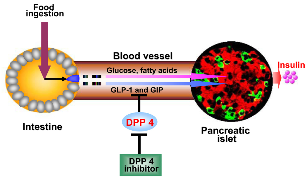
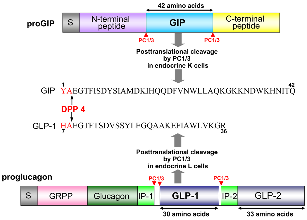
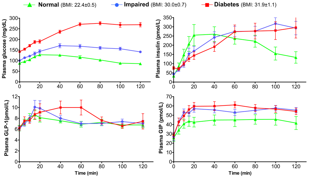
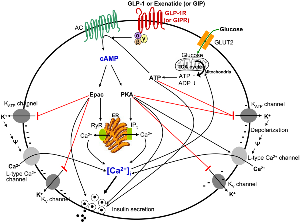
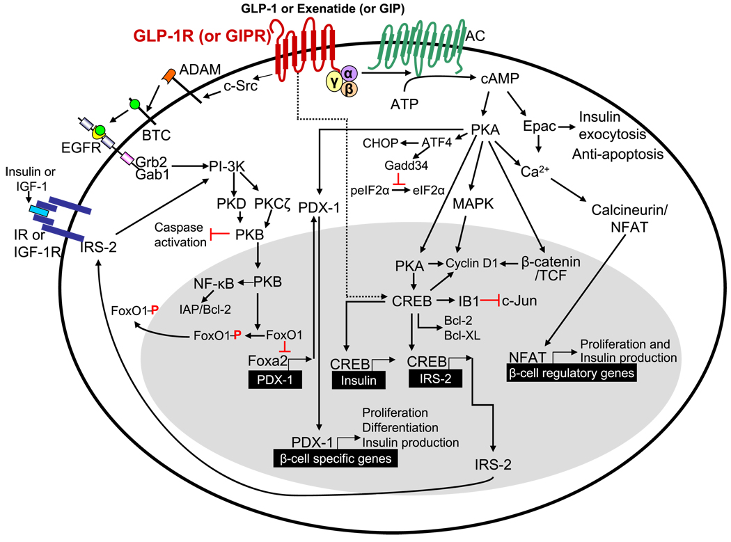
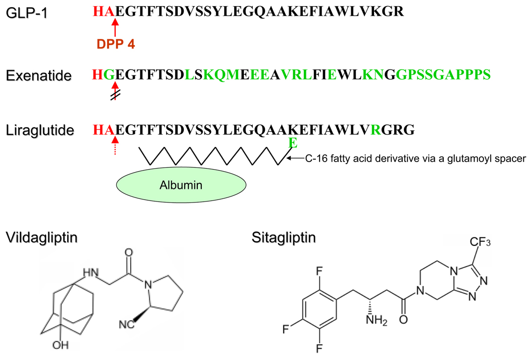
Incretins are gut hormones that are secreted from enteroendocrine cells into the blood within minutes after eating. One of their many physiological roles is to regulate the amount of insulin that is secreted after eating. In this manner, as well as others to be described in this review, their final common raison d'être is to aid in disposal of the products of digestion. There are two incretins, known as glucose-dependent insulinotropic peptide (GIP) and glucagon-like peptide-1 (GLP-1), that share many common actions in the pancreas but have distinct actions outside of the pancreas. Both incretins are rapidly deactivated by an enzyme called dipeptidyl peptidase 4 (DPP4). A lack of secretion of incretins or an increase in their clearance are not pathogenic factors in diabetes. However, in type 2 diabetes (T2DM), GIP no longer modulates glucose-dependent insulin secretion, even at supraphysiological (pharmacological) plasma levels, and therefore GIP incompetence is detrimental to beta-cell function, especially after eating. GLP-1, on the other hand, is still insulinotropic in T2DM, and this has led to the development of compounds that activate the GLP-1 receptor with a view to improving insulin secretion. Since 2005, two new classes of drugs based on incretin action have been approved for lowering blood glucose levels in T2DM: an incretin mimetic (exenatide, which is a potent long-acting agonist of the GLP-1 receptor) and an incretin enhancer (sitagliptin, which is a DPP4 inhibitor). Exenatide is injected subcutaneously twice daily and its use leads to lower blood glucose and higher insulin levels, especially in the fed state. There is glucose-dependency to its insulin secretory capacity, making it unlikely to cause low blood sugars (hypoglycemia). DPP4 inhibitors are orally active and they increase endogenous blood levels of active incretins, thus leading to prolonged incretin action. The elevated levels of GLP-1 are thought to be the mechanism underlying their blood glucose-lowering effects.


Figures

FIG. 1
FIG. 1
Schematic representation of incretin secretion and action. GIP and GLP-1 are secreted after food ingestion, and they then stimulate glucose-dependent insulin secretion. Once released, GIP and GLP-1 are subject to degradation by DPP4 on lymphocytes and on endothelial cells of blood vessels. The red cells in the islets are insulin-containing (β) cells and the green cells are glucagon-containing (α) cells.
FIG. 2
FIG. 2
Schematic representation of proGIP and proglucagon. GIP is a single 42-amino acid peptide derived from the post-translational processing of proGIP by PC1/3 in enteroendocrine K cells. It is the only functional proGIP product in all species examined to date, and there is a greater than 90% amino acid identity between human, rat, murine, porcine, and bovine sequences. GLP-1 is a post-translational cleavage product of the proglucagon gene by PC1/3 in enteroendocrine L cells and GLP-1 (7–36) amide is a major form of circulating biologically active GLP-1 in humans. In mammals, proglucagon is expressed in pancreas, enteroendocrine L cells, brain, and taste cells with an identical mRNA transcript in each tissue. Fish and bird proglucagon mRNA in pancreas and liver, however, have different 3′-ends because of differential splicing upon pancreatic expression. The Xenopus laevis proglucagon gene encodes three unique GLP-1-like peptides, each with insulinotropic properties that are capable of activating the human GLP-1R, and one of which seems more potent than human GLP-1 (Irwin et al., 1997).
FIG. 3
FIG. 3
Schematic representation of tissue-specific post-translational processing of proglucagon in the intestinal L cell and the pancreatic α cell. PC1/3 is responsible for the processing of proglucagon in the L cell to release GLP-1 (and GLP-2), and PC2, in conjunction with 7B2, is responsible for the pancreatic α cell-specific processing of proglucagon. Processing of proglucagon also occurs in brain. In taste cells of the tongue, both PC1/3 and PC2 (as well as 7B2) are present, and consequently GLP-1, GLP-2, and glucagon are all present.
FIG. 4
FIG. 4
Hormone responses to oral glucose (75 g) in nondiabetic (green), glucose intolerant (blue), and newly diagnosed type 2 diabetic (red, T2DM) subjects within Baltimore Longitudinal Study of Aging: the impaired and diabetes subjects were matched for body mass index. These subjects were not taking any glucose-lowering medications. It is evident that glucose-mediated incretin secretion (GIP and GLP-1) is not deficient in newly diagnosed T2DM.
FIG. 5
FIG. 5
A schematic representation of the main molecular events during incretin-induced insulin secretion from a β-cell. The binding of incretin ligands or agonists to the incretin receptors results in production of cAMP via adenylyl cyclase (AC) activation and subsequent activation of PKA and the Epac family of cAMP-regulated guanine nucleotide exchange factors (cAMP-GEFs), which leads to elevation of intracellular Ca2+ levels via a depolarization of plasma membrane by inhibition of KATP and KV channels after ATP generation from glucose and consequent opening of voltage-gated L-type Ca2+ channels. Intracellular Ca2+ levels are further increased via stimulation of IP3R and RyR on the ER. Long-term GLP-1 treatment also stimulates the expression of GLUT2 transporter and glucokinase (the β-cell glucose sensor), which lead to increased mitochondrial ATP synthesis. In addition, L-type Ca2+ channels are phosphorylated by PKA, resulting in an increase of their open probability and thus facilitation of enhanced Ca2+ influx. The changes in intracellular Ca2+ concentrations lead to fusion of insulin-containing vesicles to the plasma membrane and subsequent rapid exocytosis of insulin from β cells. In addition, the exocytosis of insulin-containing vesicles is directly regulated by PKA and Epac2 via interaction with the regulators of exocytosis and ATP.
FIG. 6
FIG. 6
Schema outlining the incretin downstream signal transduction pathways in a β cell. GLP-1R activation (and GIPR activation, to some extent, but this has not been as well studied as GLP-1R activation) recruits signaling mechanisms that considerably overlap, leading to promotion of β-cell proliferation and prevention of β-cell apoptosis. Dashed line indicates mechanism that is not fully delineated.
FIG. 7
FIG. 7
Structure of native GLP-1, exenatide, liraglutide, sitagliptin, and vildagliptin. Green letters indicate changes introduced in derivatives or occur naturally in exendin-4 (and replicated in the synthetic version, exenatide). The N-terminal dipeptide “HA” of GLP-1 and liraglutide is the site of proteolytic cleavage of DPP4. A broken red arrow indicates absent DPP4 activity, and a dotted red arrow indicates reduced DPP4 activity.

References

    1. Abbott CR, Monteiro M, Small CJ, Sajedi A, Smith KL, Parkinson JR, Ghatei MA, Bloom SR. The inhibitory effects of peripheral administration of peptide YY(3–36) and glucagon-like peptide-1 on food intake are attenuated by ablation of the vagal-brainstem-hypothalamic pathway. Brain Res. 2005;1044:127–131. - PubMed
    1. Acitores A, González N, Sancho V, Valverde I, Villanueva-Peñacarrillo ML. Cell signalling of glucagon-like peptide-1 action in rat skeletal muscle. J Endocrinol. 2004;180:389–398. - PubMed
    1. Agersø H, Jensen LB, Elbrønd B, Rolan P, Zdravkovic M. The pharmacokinetics, pharmacodynamics, safety and tolerability of NN2211, a new long-acting GLP-1 derivative, in healthy men. Diabetologia. 2002;45:195–202. - PubMed
    1. Ahrén B. Sensory nerves contribute to insulin secretion by glucagon-like peptide-1 in mice. Am J Physiol Regul Integr Comp Physiol. 2004;286:R269–F272. - PubMed
    1. Alcántara AI, Morales M, Delgado E, López-Delgado MI, Clemente F, Luque MA, Malaisse WJ, Valverde I, Villanueva-Peñacarrillo ML. Exendin-4 agonist and exendin(9–39)amide antagonist of the GLP-1(7–36)amide effects in liver and muscle. Arch Biochem Biophys. 1997;341:1–7. - PubMed

Publication types