. 2009 Feb;296(2):G414-23.
doi: 10.1152/ajpgi.90340.2008. Epub 2008 Dec 12.

Gastrin activates paracrine networks leading to induction of PAI-2 via MAZ and ASC-1

Affiliations

Gastrin activates paracrine networks leading to induction of PAI-2 via MAZ and ASC-1

Simon Almeida-Vega et al. Am J Physiol Gastrointest Liver Physiol. 2009 Feb.

Abstract

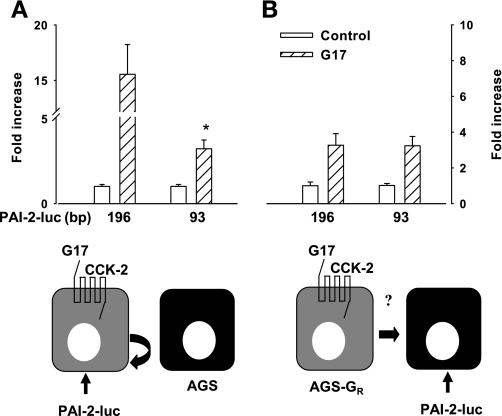
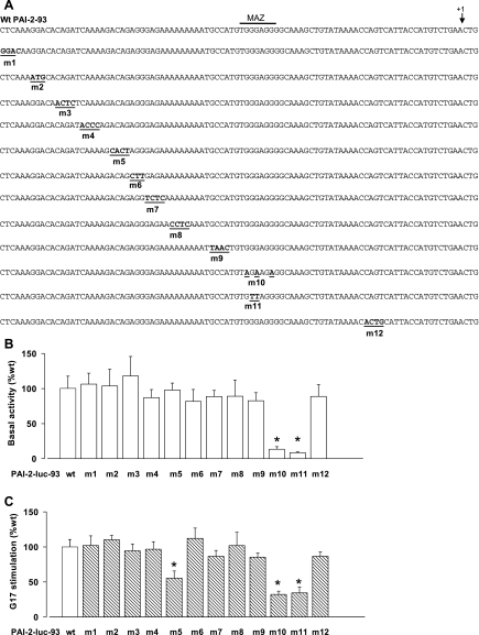
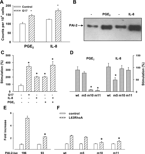
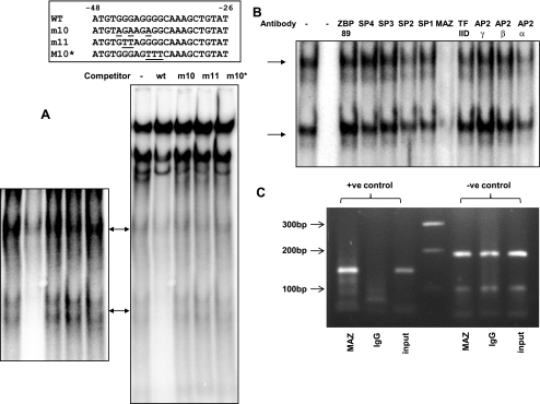
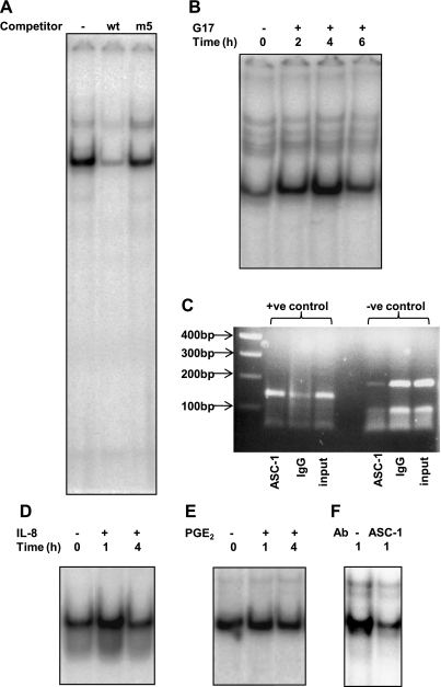
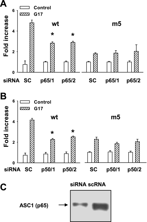
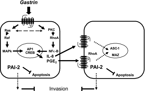
The gastric hormone gastrin regulates the expression of a variety of genes involved in control of acid secretion and also in the growth and organization of the gastric mucosa. One putative target is plasminogen activator inhibitor-2 (PAI-2), which is a component of the urokinase activator system that acts extracellularly to inhibit urokinase plasminogen activator (uPA) and intracellularly to suppress apoptosis. Previous studies have demonstrated that gastrin induces PAI-2 both in gastric epithelial cells expressing the gastrin (CCK-2) receptor and, via activation of paracrine networks, in adjacent cells that do not express the receptor. We have now sought to identify the response element(s) in the PAI-2 promoter targeted by paracrine mediators initiated by gastrin. Mutational analysis identified two putative response elements in the PAI-2 promoter that were downstream of gastrin-activated paracrine signals. One was identified as a putative MAZ site, mutation of which dramatically reduced both basal and gastrin-stimulated responses of the PAI-2 promoter by a mechanism involving PGE(2) and the small GTPase RhoA. Yeast one-hybrid screening identified the other as binding the activating signal cointegrator-1 (ASC-1) complex, which was shown to be the target of IL-8 released by gastrin. RNA interference (RNAi) knockdown of two subunits of the ASC-1 complex (p50 and p65) inhibited induction of PAI-2 expression by gastrin. The data reveal previously unsuspected transcriptional mechanisms activated as a consequence of gastrin-triggered paracrine networks and emphasize the elaborate and complex cellular control mechanisms required for a key component of tissue responses to damage and infection.


Figures

Fig. 1.
Fig. 1.
For paracrine-mediated effects of gastrin, 93 bp of the plasminogen activator inhibitor-2 (PAI-2) promoter is sufficient. A: in cells expressing the CCK-2 receptor and transfected with PAI-2-luc vectors, there was a reduction in luciferase activity in response to G17 (1 nM, 8 h) of the 93-bp PAI-2 luciferase construct compared with the 196-bp construct, *P < 0.05, t-test, n = 3. B: in contrast, when the 93-bp construct was transfected into cells not expressing CCK-2 receptors, but cocultured with cells expressing this receptor, the response to G17 (attributable to the action of paracrine mediators) was similar for both constructs and was comparable to the response to the 93-bp construct according to the protocol illustrated in A. Schematic cartoons underneath A and B illustrate the experimental design in each case (see Fig. 7 for a summary of the relevant signaling pathways). Note that in A, AGS cells not expressing the CCK-2 receptor and not transfected with PAI-2-luc vectors are included to ensure that total cell numbers were the same in the 2 conditions; in these circumstances PAI-2-luc responses are expected to reflect either direct effects of gastrin or effects consequent on activation of autocrine signaling pathways.
Fig. 2.
Fig. 2.
Mutation of the proximal 93-bp promoter region indicates that the paracrine-mediated actions of gastrin target multiple sites. A: the series of mutants studied included those with deletion of putative MAZ site (m10-11) and the GACAGA sequence (m5). The consensus MAZ binding site is indicated by a horizontal bar and the start of transcription by an arrow. B: basal expression of the wild-type (wt) and m1 to m12 promoter-reporter constructs. Results are expressed as relative to the basal luciferase activity of the wt PAI-2-93-luc construct; note the effect of m10 and m11 on basal expression. C: responses to G17 (1 nM, 8 h) of the wt and mutant promoter-reporter constructs. Results are expressed relative to the response of wt PAI-2-93-luc. *P < 0.05, ANOVA; n = 3.
Fig. 3.
Fig. 3.
Effects of PGE2, IL-8, and RhoA on wt and mutant PAI-2 promoter constructs. A: flow cytometric analysis shows that gastrin increased COX-2 and IL-8 in AGS cells expressing the CCK-2 receptor. B: Western blot shows that the abundance of wt PAI-2 protein is increased in AGS cells treated with PGE2 and IL-8. C: PGE2 (28 μM, 6 h) and IL-8 (125 ng/ml, 6 h) stimulated expression of wt PAI-2-93-luc. D: responses to PGE-2 are significantly reduced in m10 and m11 but not m5, whereas the response to IL-8 is reduced in m5 but not in m10 and m11. E: cotransfection of AGS cells with L63RhoA (0.5 μg/well) stimulates PAI-2-93-luc to a lesser extent than PAI-2-196 (*P < 0.05, t-test, n = 3). F: responses to L63RhoA of PAI-2-93-luc were significantly reduced by mutations of the MAZ site (m10-11) but not of the GACAGA site (m5). *P < 0.05, ANOVA; n = 3.
Fig. 4.
Fig. 4.
Analysis of the MAZ binding site. A: wt and mutant MAZ double-stranded (ds) oligonucleotides (sequences inset, 100× excess) were used to compete with radiolabeled wt probe binding to nuclear extracts from G17-stimulated AGS-GR cells. MAZ site-specific binding complexes are indicated by arrows. Left, whole gel; right, enhancement of MAZ site-specific complexes. B: effects of transcription factor antibodies on wt probe binding to nuclear extracts from G17-stimulated AGS-GR cells. Lane 1, no antibody, lane 2, no extract, lanes 3-12, antibodies as indicated. C: PCR analysis of chromatin immunoprecipitation (ChIP) assay. Lane 1, positive (+ve) control primers (143-bp amplicon) with MAZ immunoprecipitated DNA as template. Lane 2, positive control primers with IgG immunoprecipitated DNA as template. Lane 3, positive control primers with unprecipitated input DNA as template. Lane 4, 100-bp marker. Lanes 5-7, templates as for lanes 1-3 but with negative (−ve) control primers (186-bp amplicon).
Fig. 5.
Fig. 5.
Analysis of the −81 to −59 region of the PAI-2 promoter. A: wt and mutant ds oligonucleotides (100× excess) were used to compete with radiolabeled wt probe binding to nuclear extracts from gastrin-stimulated AGS-GR cells. B: time course of wt probe binding to nuclear extracts from gastrin-stimulated AGS-GR cells. Lane 1: unstimulated cells; lanes 2-4: cells stimulated with G17 (1 nM) for times indicated. Representative data from 3 replicate experiments. C: PCR analysis of ChIP assay. Lane 1, 100-bp marker. Lane 2, positive control primers (143-bp amplicon) with ASC-1 immunoprecipitated DNA as template. Lane 3, positive control primers with IgG immunoprecipitated DNA as template. Lane 4, positive control primers with unprecipitated input DNA as template. Lanes 5-7, templates as for lanes 2-4 but with negative control primers (186-bp amplicon). D: time course of wt probe binding to nuclear extracts from gastrin-stimulated AGS-GR cells. Lane 1, unstimulated cells; lanes 2-3, cells stimulated with IL-8 (125 ng/ml) for times indicated. E: time course of wt probe binding to nuclear extracts from gastrin-stimulated AGS-GR cells. Lane 1: unstimulated cells; lanes 2-3: cells stimulated with PGE-2 (28 μM) for the times indicated. F: effects of ASC-1 antibody on wt probe binding to nuclear extracts from G17-stimulated AGS-GR cells. Lane 1, control rabbit IgG; lane 2, ASC-1 antibody.
Fig. 6.
Fig. 6.
Knockdown of the ASC-1 complex inhibits luciferase activity of the wt 93-bp promoter but not the m5 mutant. A: 2 independent small interfering RNAs (siRNAs) to p65 subunit (p65/1 and p65/2, 30 nM, 48 h) significantly reduce luciferase expression of the wt but not m5 PAI-2-93-luc, compared with scrambled control siRNA (SC, scRNA). B: 2 siRNAs to p50 subunit (p50/1 and p50/2, 30 nM, 48 h) also inhibit luciferase expression of the wt but not the m5 PAI-2-93-luc construct. C: Western blot shows depletion of ASC-1 protein by siRNA treatment compared with scRNA. *P < 0.05, ANOVA; n = 3.
Fig. 7.
Fig. 7.
Schematic representation of regulation of PAI-2 expression by gastrin. Gastrin regulates PAI-2 expression in both CCK-2 receptor-expressing cells by acting directly through its receptor (left) and in neighboring cells, via paracrine mediators released from CCK-2 receptor expressing cells (right). Direct regulation involves the CRE and AP-1 sites via PKC, Ras, Raf, RhoA, and the NF-κB pathways. The paracrine mediators IL-8 and PGE2 are released in response to gastrin stimulation; IL-8 acts through a GACAGA site via the ASC-1 complex, whereas PGE2 targets the MAZ site via the small GTPase RhoA.

References

    1. Andreasen PA, Egelund R, Petersen HH. The plasminogen activation system in tumor growth, invasion, and metastasis. Cell Mol Life Sci 57: 25–40, 2000. - PMC - PubMed
    1. Andreasen PA, Kjoller L, Christensen L, Duffy MJ. The urokinase-type plasminogen activator system in cancer metastasis: a review. Int J Cancer 72: 1–22, 1997. - PubMed
    1. Ansorge N, Juttner S, Cramer T, Schmidt WE, Hocker M, Schmitz F. An upstream CRE-E-box element is essential for gastrin-dependent activation of the cyclooxygenase-2 gene in human colon cancer cells. Regul Pept 144: 25–33, 2007. - PubMed
    1. Antalis TM, Costelloe E, Muddiman J, Ogbourne S, Donnan K. Regulation of the plasminogen activator inhibitor type-2 gene in monocytes: localization of an upstream transcriptional silencer. Blood 88: 3686–3697, 1996. - PubMed
    1. Backhed F, Torstensson E, Seguin D, Richter-Dahlfors A, Rokbi B. Helicobacter pylori infection induces interleukin-8 receptor expression in the human gastric epithelium. Infect Immun 71: 3357–3360, 2003. - PMC - PubMed

Publication types

MeSH terms