Comparative Study
. 2009 Apr 30;113(18):4391-402.
doi: 10.1182/blood-2008-09-178228. Epub 2008 Dec 12.

microRNA expression in the biology, prognosis, and therapy of Waldenström macroglobulinemia

Affiliations
Comparative Study

microRNA expression in the biology, prognosis, and therapy of Waldenström macroglobulinemia

Aldo M Roccaro et al. Blood. .

Abstract

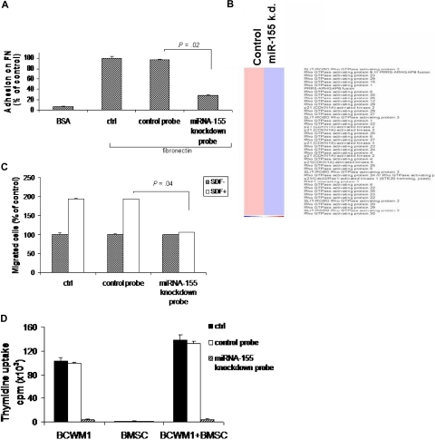
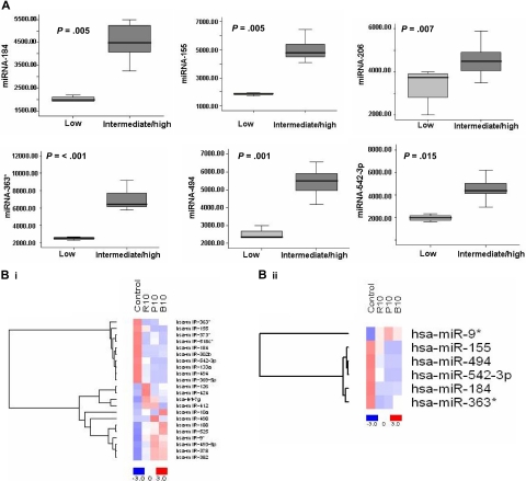
Multilevel genetic characterization of Waldenström macroglobulinemia (WM) is required to improve our understanding of the underlying molecular changes that lead to the initiation and progression of this disease. We performed microRNA-expression profiling of bone marrow-derived CD19(+) WM cells, compared with their normal cellular counterparts and validated data by quantitative reverse-transcription-polymerase chain reaction (qRT-PCR). We identified a WM-specific microRNA signature characterized by increased expression of microRNA-363*/-206/-494/-155/-184/-542-3p, and decreased expression of microRNA-9* (ANOVA; P < .01). We found that microRNA-155 regulates proliferation and growth of WM cells in vitro and in vivo, by inhibiting MAPK/ERK, PI3/AKT, and NF-kappaB pathways. Potential microRNA-155 target genes were identified using gene-expression profiling and included genes involved in cell-cycle progression, adhesion, and migration. Importantly, increased expression of the 6 miRNAs significantly correlated with a poorer outcome predicted by the International Prognostic Staging System for WM. We further demonstrated that therapeutic agents commonly used in WM alter the levels of the major miRNAs identified, by inducing downmodulation of 5 increased miRNAs and up-modulation of patient-downexpressed miRNA-9*. These data indicate that microRNAs play a pivotal role in the biology of WM; represent important prognostic marker; and provide the basis for the development of new microRNA-based targeted therapies in WM.


Figures

Figure 1
Figure 1
microRNA expression pattern in WM and healthy donors. (A) miRNA analysis has been performed on total RNA isolated from BM CD19+ WM cells, normal bone marrow (NBM)– and peripheral blood (PBMC)–derived CD19+ counterparts, and WM cell line (BCWM.1). Heat map was generated after unsupervised hierarchic cluster analysis of all unfiltered data. Differential expression of miRNA is shown by the intensity of red (up-regulation) versus blue (down-regulation). (B) Supervised hierarchic clustering analysis was performed using ANOVA test. Differential expression of miRNA patterns is shown by the intensity of red (up-regulation) versus blue (down-regulation). (C) Differential distribution of each indicated miRNA in all WM patients (WM) compared with healthy donors (HDs): mean values were compared using Mann-Whitney U rank sum test; bars indicate standard errors; P value (*) for each miRNA is indicated.
Figure 2
Figure 2
Validation of miRNA expression levels by qRT-PCR. The amount of miRNA-184, -206, -494, -363*, -155, 542-3p, and -9* in WM samples compared with normal controls, evaluated by qRT-PCR (□) and microbead miRNA profiling (■). Results are expressed as fold change of the miRNA expression in bone marrow WM CD19+ cells with respect to average miRNA expression from 3 healthy donors' bone marrow CD19+ cells (BM). qRT-PCR data were obtained using the ΔΔCt method, with normalization to the reference RNU6B microRNA. Bars represent SD.
Figure 3
Figure 3
miRNA-155 modulates proliferation and cell cycle of WM cells. (A,B) WM cells (miRNA-155 knockdown probe–transfected, control probe–transfected BCWM.1 cells) were harvested at 24 and 48 hours after transfection; DNA synthesis and cytotoxicity were assessed by thymidine uptake and MTT assays, respectively. Nontransfected BCWM.1 cells were used as controls (ctrl). *P < .001; **P < .001. (C) Cells were first arrested and synchronized in G2/M phase by growth in 80 nM nocodazole for 16 hours. Cells were then washed and regrown using fresh media. After 6 hours, cell-cycle analysis was performed by propidium iodide staining. (D) Purified cRNA (15 μg) isolated from WM cells (control probe–transfected, miRNA-155 knockdown probe–transfected BCWM.1 cells) was hybridized to HG-U133Plus2.0 GeneChip (Affymetrix). (i) Up-regulation of cyclin-dependent kinase inhibitors and down-regulation of cyclins and cyclin-dependent kinases. (ii) Up-regulation of p53 and p53 family members and down-regulation of p53 negative regulators. Fold change is shown by the intensity of induction (red) or suppression (blue). (E) BCWM.1 cells (miRNA-155 knockdown probe transfected, control probe transfected) were harvested at 8 hours after transfection. Whole-cell lysates were subjected to Western blotting using anti–phospho (p)-ERK, –p-AKT, -AKT, –p-GSK3α/β, –p-S6R, and -actin antibodies; nontransfected BCWM.1 cells were used as controls (ctrl). (F) BCWM.1 cells (miRNA-155 knockdown probe transfected, control probe transfected) were harvested at 8 hours after transfection and treated with and without TNF-α (10 ng/mL) for 20 minutes; nontransfected BCWM.1 cells were used as control (ctrl). NF-κBp65 transcription factor binding to its consensus sequence on the plate-bound oligonucleotide was measured in nuclear extracts. Wild-type and mutant are wild-type and mutated consensus competitor oligonucleotides, respectively. All results represent means (±] SD) of triplicate experiments. (G) BCWM.1 cells (miRNA-155 knockdown probe transfected, control probe transfected) were harvested at 8 hours after transfection and treated with and without TNF-α (10 ng/mL) for 20 minutes; nontransfected BCWM.1 cells were used as control (ctrl). Immunocytochemical analysis was assessed using anti–p-NF-κBp65 antibody, with DAPI used to stain nuclei.
Figure 4
Figure 4
miRNA-155 knockdown inhibited WM cells in the context of bone marrow microenvironment in vitro. WM cells (miRNA-155 knockdown probe–transfected, control probe–transfected BCWM.1 cells) were harvested at 24 hours after transfection. Nontransfected BCWM.1 cells were used as control (ctrl). (A) Adhesion assay to fibronectin (FN). Control probe–transfected and nontransfected cells showed significant increase in adhesion to FN, compared with BSA, used as control. miRNA-155 knockdown probe–transfected cells showed lower adhesion to FN. (B) Purified cRNA (15 μg) isolated from WM cells (control probe–transfected, miRNA-155 knockdown probe–transfected BCWM.1 cells) was hybridized to HG-U133Plus2.0 GeneChip (Affymetrix). Down-regulation of several genes involved in the adhesion process. Fold change is shown by the intensity of induction (red) or suppression (blue). (C) Transwell migration assay. SDF-1 (30 nM) was placed in the lower chambers and migration was determined after 4 hours. Control probe–transfected and nontransfected cells showed significant migration with SDF-1 30 nM, whereas the miRNA-155 knockdown probe–transfected cells showed minimal migration in response to SDF-1. (D) BCWM.1 cells were cultured for 48 hours in the presence or absence of BMSCs, and cell proliferation was assessed using [3H]-thymidine uptake assay. All data represent mean (±SD) of triplicate experiments.
Figure 5
Figure 5
miRNA-155 knockdown targets WM cells in the context of bone marrow microenvironment in vivo. (A) In vivo confocal imaging. GFP+ WM cells were transfected using either control probe or miRNA-155 knockdown probe, and then injected in mice 24 hours after transfection. GFP+ WM cells (green) were excited with a 491-nm solid state laser. Blood vessels (red) were imaged using Angiosense-750 (VisEn Medical) excited with a 750-nm laser. Images of parasagittal vasculature and GFP+ tumor cells in the mouse skull bone marrow. Control probe–transfected cells homed and adhered more than the miRNA-155–transfected counterparts. (B) Detection of human IgM from serum of control probe–injected (n = 4) and miRNA-155 knockdown probe–injected (n = 4) mice. Serum of noninjected mice was used as negative control. Average obtained from each group is shown. (C) Kaplan-Meier curve showing survival in each group; mean survival was 14 days versus 20 days (P < .001), and overall survival was 15 days versus 21 days, respectively, in control probe–injected versus miRNA-155 knockdown probe–injected mice.
Figure 6
Figure 6
Differentially expressed miRNAs are associated with prognosis in WM patients, and drugs used to treat WM modulate their expression level. (A) Significant differential distribution of each indicated miRNA among subgroups of WM patients with differential clinical-prognostic features (low risk versus intermediate/high risk). Mean values were compared using Mann-Whitney U rank sum test; bars indicate standard errors. P value for each miRNA is indicated. (B) BCWM1 cells were treated with either rituximab (R; 10 μg/mL; 2 hours), perifosine (P; 10 μM; 6 hours), or bortezomib (B; 10 nM; 6 hours), and miRNA analysis was performed on total RNA isolated from treated and untreated cells. Heat map was generated after supervised hierarchic cluster analysis and comparison among the 2 groups (treated vs untreated samples). Differential expression of miRNA is shown by the intensity of red (up-regulation) or blue (down-regulation).

Comment in

Similar articles

Cited by

References

    1. Lee RC, Feinbaum RL, Ambros V. The C-Elegans heterochronic gene Lin-4 encodes small RNAs with antisense complementarity to Lin-14. Cell. 1993;75:843–854. - PubMed
    1. Bentwich I, Avniel A, Karov Y, et al. Identification of hundreds of conserved and nonconserved human microRNAs. Nat Genet. 2005;37:766–770. - PubMed
    1. Xie X, Lu J, Kulbokas EJ, et al. Systematic discovery of regulatory motifs in human promoters and 3′UTRs by comparison of several mammals. Nature. 2005;434:338–345. - PMC - PubMed
    1. He L, Hannon GJ. MicroRNAs: small RNAs with a big role in gene regulation. Nat Rev Genet. 2004;5:522–531. - PubMed
    1. Roldo C, Missiaglia E, Hagan JP, et al. MicroRNA expression abnormalities in pancreatic endocrine and acinar tumors are associated with distinctive pathologic features and clinical behaviour. J Clin Oncol. 2006;24:4677–4684. - PubMed

Publication types

MeSH terms