

Retinal ion regulation in a mouse model of diabetic retinopathy: natural history and the effect of Cu/Zn superoxide dismutase overexpression
- PMID: 19074809
- PMCID: PMC2688071
- DOI: 10.1167/iovs.08-2918
Retinal ion regulation in a mouse model of diabetic retinopathy: natural history and the effect of Cu/Zn superoxide dismutase overexpression
Abstract
Purpose: To test the hypotheses that manganese-enhanced MRI (MEMRI) is useful in evaluating intraretinal ion dysregulation in wild-type (WT) and Cu/Zn superoxide dismutase (SOD1) overexpressor mice.
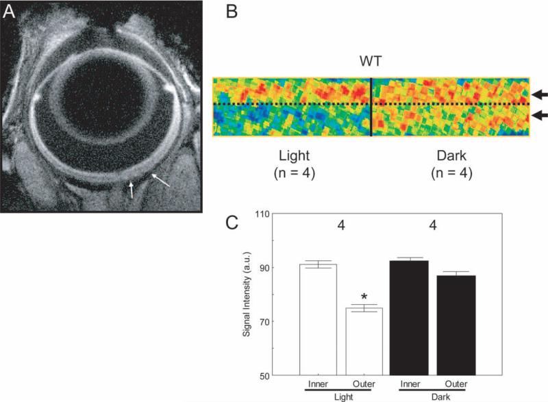
Methods: Central intraretinal ion activity and retinal thickness were measured from high-resolution data of light- and dark-adapted WT C57BL/6 mice (to gauge MEMRI sensitivity to normal visual processing in mice) and dark-adapted diabetic and nondiabetic WT and Cu/Zn superoxide dismutase overexpressor (SOD1OE) mice. Glycated hemoglobin and retinal vascular histopathology were also determined.
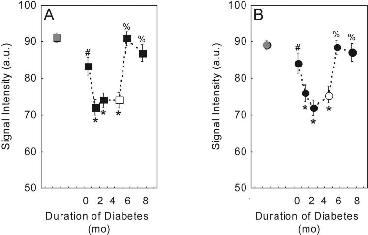
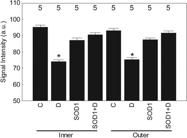
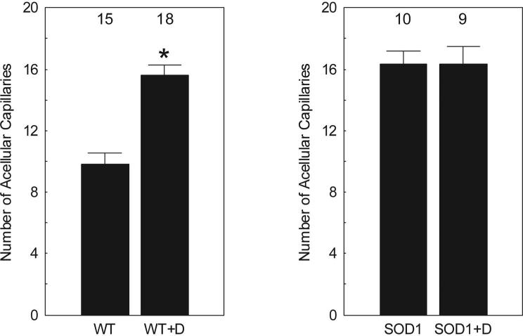
Results: In WT mice, light adaptation reduced outer retinal manganese uptake compared with that in dark adaptation; no effect on inner retinal uptake was found. In diabetic WT mice, intraretinal manganese uptake became subnormal between 1.5 and 4 months of diabetes onset and then relatively increased. Central retinal thickness, as determined with MEMRI, decreased as a function of age in diabetic mice but remained constant in control mice. Nondiabetic SOD1OE mice had normal retinal manganese uptake but subnormal retinal thickness and supernormal acellular capillary density. At 4.2 months of diabetes, SOD1OE mice had normal manganese uptake and no further thinning; acellular capillaries frequency did not increase by 9 to 10 months of diabetes.
Conclusions: In emerging diabetic retinopathy, MEMRI provided an analytic measure of an ionic dysregulatory pattern that was sensitive to SOD1 overexpression. The potential benefit of SOD1 overexpression to inhibit retinal abnormality in this model is limited by the retinal and vascular degeneration that develops independently of diabetes.
Figures
References
-
- Grunwald JE, Riva CE, Brucker AJ, Sinclair SH, Petrig BL. Altered retinal vascular response to 100% oxygen breathing in diabetes mellitus. Ophthalmology. 1984;91(12):1447–1452. - PubMed
-
- Sinclair SH, Grunwald JE, Riva CE, Braunstein SN, Nichols CW, Schwartz SS. Retinal vascular autoregulation in diabetes mellitus. Ophthalmology. 1982;89(7):748–750. - PubMed
-
- Trick GL, Berkowitz BA. Retinal oxygenation response and retinopathy. Prog Retin Eye Res. 2005;24(2):259–274. - PubMed
-
- Phipps JA, Fletcher EL, Vingrys AJ. Paired-flash identification of rod and cone dysfunction in the diabetic rat. Invest Ophthalmol Vis Sci. 2004;45(12):4592–4600. - PubMed
-
- Han Y, Bearse MA, Jr, Schneck ME, Barez S, Jacobsen CH, Adams AJ. Multifocal electroretinogram delays predict sites of subsequent diabetic retinopathy. Invest Ophthalmol Vis Sci. 2004;45(3):948–954. - PubMed
Publication types
MeSH terms
Substances
Grants and funding
LinkOut - more resources
Full Text Sources
Medical
Molecular Biology Databases
Miscellaneous
