

1,1-Bis(3'-indolyl)-1-(p-chlorophenyl)methane activates the orphan nuclear receptor Nurr1 and inhibits bladder cancer growth
- PMID: 19074857
- PMCID: PMC4948978
- DOI: 10.1158/1535-7163.MCT-08-0730
1,1-Bis(3'-indolyl)-1-(p-chlorophenyl)methane activates the orphan nuclear receptor Nurr1 and inhibits bladder cancer growth
Abstract
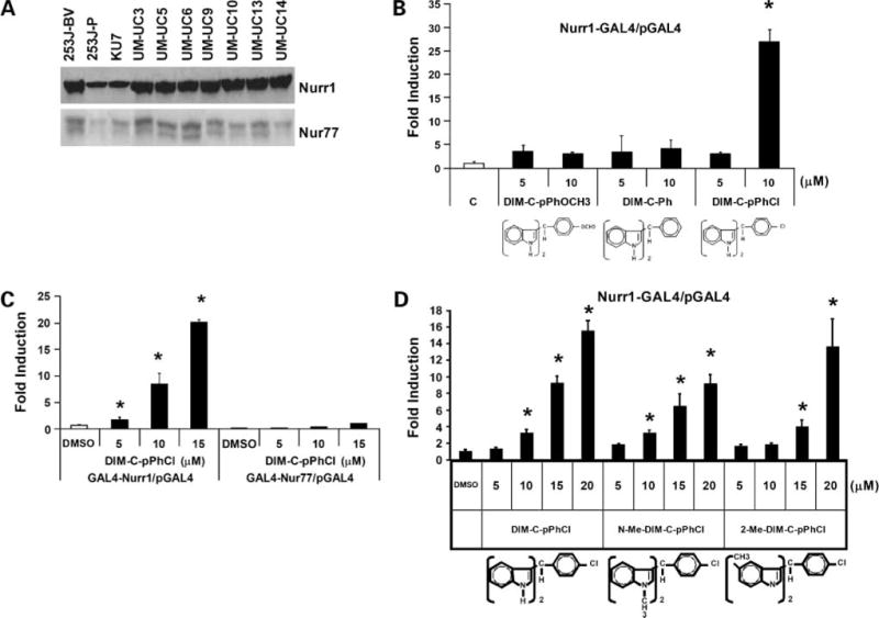
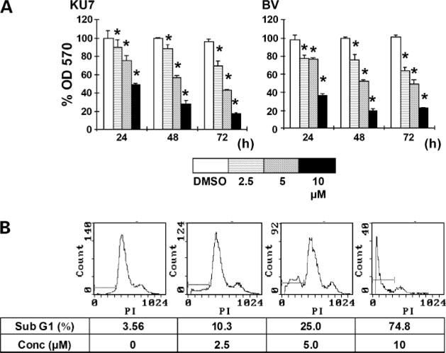
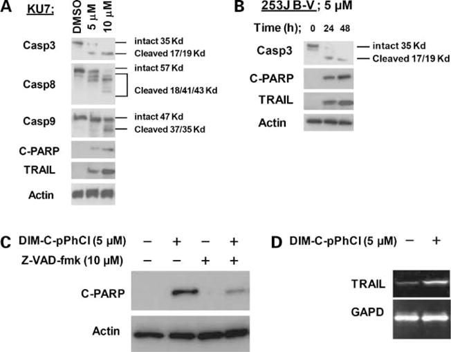
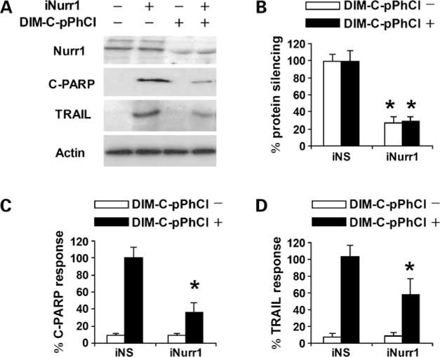
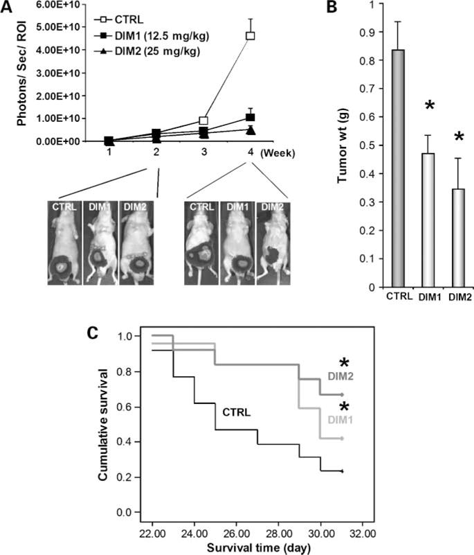
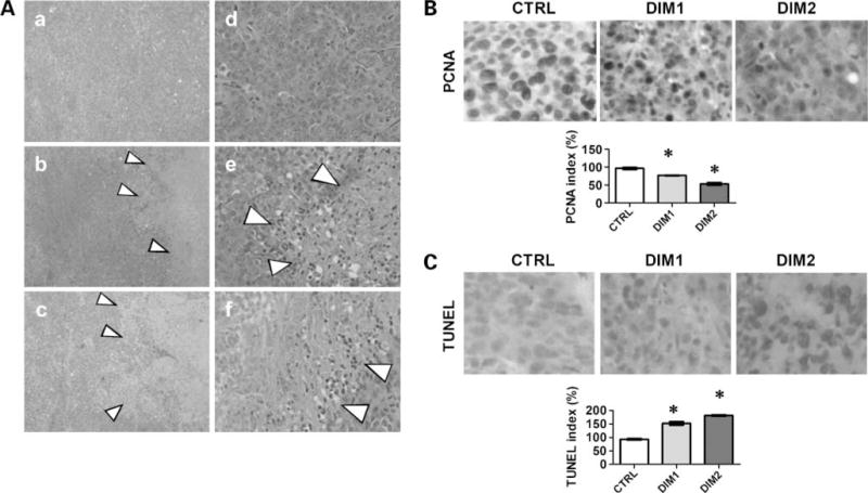
Nurr1 is an orphan nuclear receptor and a member of the nerve growth factor I-B subfamily of transcription factors with no known endogenous ligand or stimulator. We show, for the first time, evidence that Nurr1 is expressed in a panel of 11 human bladder cancer cell lines. A new class of methylene-substituted diindolylmethanes (C-DIM) were screened and 1,1-bis(3'-indolyl)-1-(p-chlorophenyl)methane (DIM-C-pPhCl) activated the ligand-binding domain of Nurr1. Treatment of bladder cancer cells with Nurr1-active C-DIM resulted in decreased cell survival (MTT assay) and induction of cell death pathways, resulting in poly(ADP-ribose) polymerase cleavage and DNA fragmentation. The specificity of the Nurr1-active compound was shown using RNA interference in 253J B-V cells, whereby small interfering RNA against Nurr1 attenuated ligand-dependent activation of Nurr1 and poly(ADP-ribose) polymerase cleavage. Furthermore, activation of Nurr1 resulted in stimulation of tumor necrosis factor-related apoptosis-inducing ligand and small interfering RNA experiments attenuated tumor necrosis factor-related apoptosis-inducing ligand production. In an orthotopic model of human bladder tumors established in nude mice, administration of a Nurr1-active C-DIM suppressed bladder cancer growth. These results identify Nurr1 as a potential target for bladder cancer therapy and also identify a novel agent for activating Nurr1.
Conflict of interest statement
A.M. Kamat: grant support from Bioniche, Abbott Molecular, Bayer, Tetralogic Pharmaceuticals, Halozyne, and Indevus; consultant to Plantacor. No other potential conflicts of interest were disclosed.
Figures
Similar articles
-
Activation of Nur77 by selected 1,1-Bis(3'-indolyl)-1-(p-substituted phenyl)methanes induces apoptosis through nuclear pathways.J Biol Chem. 2005 Jul 1;280(26):24903-14. doi: 10.1074/jbc.M500107200. Epub 2005 May 3. J Biol Chem. 2005. PMID: 15871945
-
Activation of nerve growth factor-induced B alpha by methylene-substituted diindolylmethanes in bladder cancer cells induces apoptosis and inhibits tumor growth.Mol Pharmacol. 2010 Mar;77(3):396-404. doi: 10.1124/mol.109.061143. Epub 2009 Dec 18. Mol Pharmacol. 2010. PMID: 20023005 Free PMC article.
-
Nur77 agonists induce proapoptotic genes and responses in colon cancer cells through nuclear receptor-dependent and nuclear receptor-independent pathways.Cancer Res. 2007 Jan 15;67(2):674-83. doi: 10.1158/0008-5472.CAN-06-2907. Cancer Res. 2007. PMID: 17234778
-
Structure-dependent activation of gene expression by bis-indole and quinoline-derived activators of nuclear receptor 4A2.Chem Biol Drug Des. 2019 Oct;94(4):1711-1720. doi: 10.1111/cbdd.13564. Epub 2019 Jul 21. Chem Biol Drug Des. 2019. PMID: 31102570 Free PMC article. Review.
-
Potent synthetic and endogenous ligands for the adopted orphan nuclear receptor Nurr1.Exp Mol Med. 2021 Jan;53(1):19-29. doi: 10.1038/s12276-021-00555-5. Epub 2021 Jan 21. Exp Mol Med. 2021. PMID: 33479411 Free PMC article. Review.
Cited by
-
Ultra-flexible nanocarriers for enhanced topical delivery of a highly lipophilic antioxidative molecule for skin cancer chemoprevention.Colloids Surf B Biointerfaces. 2016 Jul 1;143:156-167. doi: 10.1016/j.colsurfb.2016.03.036. Epub 2016 Mar 15. Colloids Surf B Biointerfaces. 2016. PMID: 27003466 Free PMC article.
-
A pan-cancer analysis unveiling the function of NR4A family genes in tumor immune microenvironment, prognosis, and drug response.Genes Genomics. 2024 Aug;46(8):977-990. doi: 10.1007/s13258-024-01539-1. Epub 2024 Jul 8. Genes Genomics. 2024. PMID: 38976216
-
Unveiling the methionine cycle: a key metabolic signature and NR4A2 as a methionine-responsive oncogene in esophageal squamous cell carcinoma.Cell Death Differ. 2024 May;31(5):558-573. doi: 10.1038/s41418-024-01285-7. Epub 2024 Apr 3. Cell Death Differ. 2024. PMID: 38570607 Free PMC article.
-
Pharmacological Activators of the NR4A Nuclear Receptors Enhance LTP in a CREB/CBP-Dependent Manner.Neuropsychopharmacology. 2017 May;42(6):1243-1253. doi: 10.1038/npp.2016.253. Epub 2016 Nov 11. Neuropsychopharmacology. 2017. PMID: 27834392 Free PMC article.
-
Orphan nuclear receptor Nurr1 as a potential novel marker for progression in human pancreatic ductal adenocarcinoma.Exp Ther Med. 2017 Feb;13(2):551-559. doi: 10.3892/etm.2016.3968. Epub 2016 Dec 13. Exp Ther Med. 2017. PMID: 28352330 Free PMC article.
References
-
- Jemal A, Siegel R, Ward E, et al. Cancer statistics, 2008. CA Cancer J Clin. 2008;58:71–96. - PubMed
-
- Dinney CP, McConkey DJ, Millikan RE, et al. Focus on bladder cancer. Cancer Cell. 2004;6:111–6. - PubMed
-
- Bajorin DF, Dodd PM, Mazumdar M, et al. Long-term survival in metastatic transitional-cell carcinoma and prognostic factors predicting outcome of therapy. J Clin Oncol. 1999;17:3173–81. - PubMed
-
- Steinmetz AC, Renaud JP, Moras D. Binding of ligands and activation of transcription by nuclear receptors. Annu Rev Biophys Biomol Struct. 2001;30:329–59. - PubMed
-
- Wansa KD, Harris JM, Muscat GE, et al. The activation function-1 domain of Nur77/NR4A1 mediates trans-activation, cell specificity, and coactivator recruitment. J Biol Chem. 2002;277:33001–11. - PubMed
Publication types
MeSH terms
Substances
Grants and funding
LinkOut - more resources
Full Text Sources
Other Literature Sources
Medical
Miscellaneous
