. 2008 Dec 15;68(24):10349-57.
doi: 10.1158/0008-5472.CAN-08-2754.

250K single nucleotide polymorphism array karyotyping identifies acquired uniparental disomy and homozygous mutations, including novel missense substitutions of c-Cbl, in myeloid malignancies

Affiliations

250K single nucleotide polymorphism array karyotyping identifies acquired uniparental disomy and homozygous mutations, including novel missense substitutions of c-Cbl, in myeloid malignancies

Andrew J Dunbar et al. Cancer Res. .

Abstract

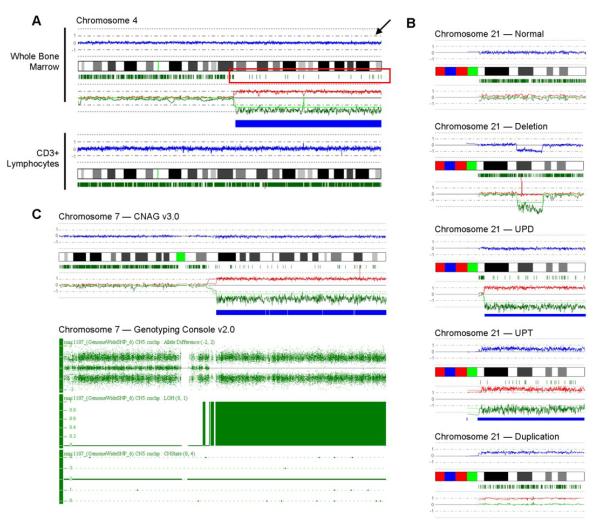
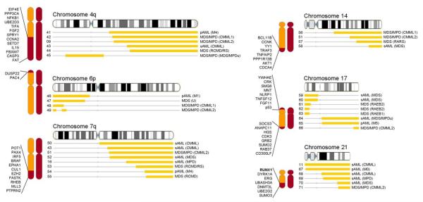
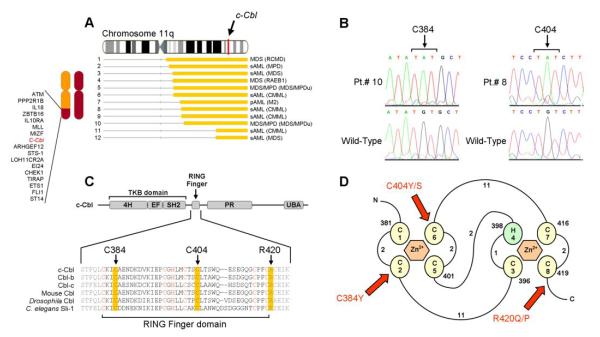
Two types of acquired loss of heterozygosity are possible in cancer: deletions and copy-neutral uniparental disomy (UPD). Conventionally, copy number losses are identified using metaphase cytogenetics, whereas detection of UPD is accomplished by microsatellite and copy number analysis and as such, is not often used clinically. Recently, introduction of single nucleotide polymorphism (SNP) microarrays has allowed for the systematic and sensitive detection of UPD in hematologic malignancies and other cancers. In this study, we have applied 250K SNP array technology to detect previously cryptic chromosomal changes, particularly UPD, in a cohort of 301 patients with myelodysplastic syndromes (MDS), overlap MDS/myeloproliferative disorders (MPD), MPD, and acute myeloid leukemia. We show that UPD is a common chromosomal defect in myeloid malignancies, particularly in chronic myelomonocytic leukemia (CMML; 48%) and MDS/MPD-unclassifiable (38%). Furthermore, we show that mapping minimally overlapping segmental UPD regions can help target the search for both known and unknown pathogenic mutations, including newly identified missense mutations in the proto-oncogene c-Cbl in 7 of 12 patients with UPD11q. Acquired mutations of c-Cbl E3 ubiquitin ligase may explain the pathogenesis of a clonal process in a subset of MDS/MPD, including CMML.


Figures

Figure 1
Figure 1. Detecting acquired, segmental UPD using 250K SNP-A technology
(A) SNP-A “karyograms” of both whole bone marrow cells and CD3+ lymphocytes in one patient demonstrate the somatic nature of acquired UPD. In this representative sample, chromosome (ch.) 4 is displayed. The blue line (indicated by the arrow) represents average copy number (CN) signal intensity of SNPs on the array chip. In this instance, there are no CN variations, and thus the blue line does not deviate from normal diploid CN. The green marks below the idiogram represent heterozygosity at particular DNA loci. In the region of UPD seen in whole bone marrow cells, drastic reduction of heterozygous loci denotes the region of UPD (red box). Remaining green marks in the region of UPD delineate the presence of non-clonal cells in the sample. The green and red lines represent the AsCNAR algorithm and show the relative signal intensity for individual homologs. In CD3+ sorted lymphocytes, a normal ch. 4 is seen. (B) SNP-A analysis of chromosome 21 in 1 healthy control and in 4 patients demonstrates the various types of lesions observed: deletions, UPD, duplications, and in a unique patient, uniparental trisomy (UPT). (C) Comparison of Affymetrix 250K and 6.0 arrays in the detection of UPD7q for one patient. CNAG v3.0 analysis (top) shows clear UPD of ch. 7 both by loss of heterozygous loci and allelic imbalance. Repeated testing on the 6.0 array and analysis using Genotyping Console v2.0 software (bottom) confirms the 250K SNP-A findings. Note that the Genotyping Console output includes Allele Difference, LOH, and CN variation (CNState) plots. The Allele Difference graph represents the genotypes for each individual SNP. Dots with a value of 1 represent SNPs with an “AA” genotype, while those with a value of -1 represent SNPs with a “BB” genotype. Dots at 0 represent heterozygous SNPs (“AB”). Complete loss of all “AB” SNPs indicates copy-neutral LOH. This is further demonstrated by both the LOH and CNState graphs, which show no loss in copy number but clear LOH.
Figure 2
Figure 2. Acquired UPD detected by 250K SNP-A may serve as a molecular marker for mutated genes in patients
Topographical maps show regions of UPD in individual patients on chromosomes 9, 13 and 1 (Left). Yellow bars corresponding to the idiogram represent the regions affected for each patient. Individual diagnoses are listed to the right of the depicted lesion (disease sub-types are indicated in parentheses; for those patients who transformed to secondary AML, the initial presentation is given in parentheses). Red lines on the idiogram represent the physical location of the JAK2, FLT3 and c-MPL genes respectively. To the right: mutational screening of JAK2, FLT3, NRAS and c-MPL in patients with segmental UPD in the region of each gene confirms their homozygous status (note that sequencing analysis of NRAS exon 2 in patient 31 appears heterozygous due to the presence of non-clonal cells in the sample). Cartoons to the left of the maps represent the diploid chromosome set and identify the minimally overlapping region on each chromosome along with representative candidate genes in the region, regardless if the segment overlaps with known gene mutations or not. Gel images have been cropped and enhanced.
Figure 3
Figure 3. Genome-wide scan of overlapping UPD in patients
Topographical maps show all overlapping regions of UPD occurring in greater than 2 patients. Gray bars corresponding to the idiogram represent the regions affected for each patient. Individual diagnoses are listed to the right of the depicted lesion. Cartoons to the left of the maps represent the diploid chromosome set and identify the minimally overlapping region on the chromosome along with representative candidate genes in each region.
Figure 4
Figure 4. Identification of unique, missense mutations in c-Cbl (11q23.3)
(A) Map of individual UPD lesions on chromosome 11q and the location of the c-Cbl gene. (B) Direct genomic DNA sequencing of exon 8 in c-Cbl reveals the presence of missense mutations. Each base pair change occurs in a homozygous state due to the copy-neutral LOH and results in the substitution of cysteine residues for tyrosine or serine residues at positions 384 (in patients 9 and 10) and 404 (in patients 6, 8, and 12). (C) Schematic representation showing the major domains of mammalian c-Cbl, primarily the tyrosine kinase binding (TKB) domain, RING finger, proline-rich (PR) region, and ubiquitin-associated domain (UBA). Also shown is the amino acid sequence spanning the RING finger domain of mammalian c-Cbl along with homologs Cbl-b, Cbl-c, and those found in Mus musculus, Drosophila, and C. elegans (Sli-1). Critical cysteine/histidine residues that make up the RING finger domain are indicated in red. Residues affected by mutations are highlighted in yellow. (D) Schematic of the prototypical RING finger domain found in c-Cbl (adapted from (39) (Fig1)) C’s denote cysteine residues; H, histidine residues and are numbered in the order with which they occur in the domain. Curved lines represent amino acid chains and are accompanied by numbers which denote their lengths. Red arrows indicate the affected residues in patients positive for the mutations and are accompanied by the resulting amino acid substitutions.

References

    1. Robinson WP. Mechanisms leading to uniparental disomy and their clinical consequences. Bioessays. 2000;22:452–9. - PubMed
    1. Jiang YH, Bressler J, Beaudet AL. Epigenetics and human disease. Annu.Rev.Genomics Hum.Genet. 2004;5:479–510. - PubMed
    1. Kralovics R, Guan Y, Prchal JT. Acquired uniparental disomy of chromosome 9p is a frequent stem cell defect in polycythemia vera. Exp.Hematol. 2002;30:229–36. - PubMed
    1. Baxter EJ, Scott LM, Campbell PJ, et al. Acquired mutation of the tyrosine kinase JAK2 in human myeloproliferative disorders. Lancet. 2005;365:1054–61. - PubMed
    1. Kralovics R, Passamonti F, Buser AS, et al. A gain-of-function mutation of JAK2 in myeloproliferative disorders. N Engl J Med. 2005;352:1779–90. - PubMed

Publication types