. 2009 Feb;37(3):804-14.
doi: 10.1093/nar/gkn999. Epub 2008 Dec 15.

The crystal structure of a replicative hexameric helicase DnaC and its complex with single-stranded DNA

Affiliations

The crystal structure of a replicative hexameric helicase DnaC and its complex with single-stranded DNA

Yu-Hua Lo et al. Nucleic Acids Res. 2009 Feb.

Abstract

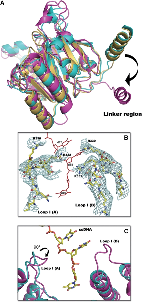
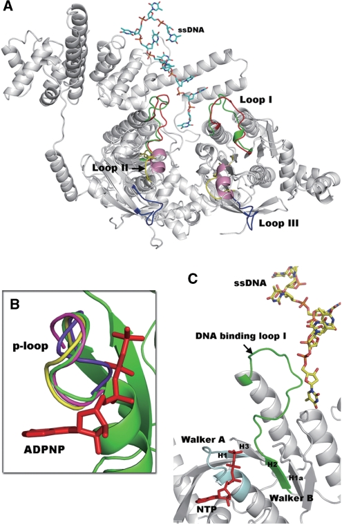
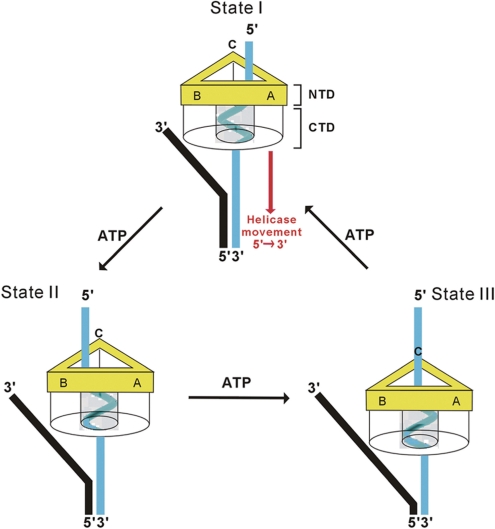
DNA helicases are motor proteins that play essential roles in DNA replication, repair and recombination. In the replicative hexameric helicase, the fundamental reaction is the unwinding of duplex DNA; however, our understanding of this function remains vague due to insufficient structural information. Here, we report two crystal structures of the DnaB-family replicative helicase from Geobacillus kaustophilus HTA426 (GkDnaC) in the apo-form and bound to single-stranded DNA (ssDNA). The GkDnaC-ssDNA complex structure reveals that three symmetrical basic grooves on the interior surface of the hexamer individually encircle ssDNA. The ssDNA-binding pockets in this structure are directed toward the N-terminal domain collar of the hexameric ring, thus orienting the ssDNA toward the DnaG primase to facilitate the synthesis of short RNA primers. These findings provide insight into the mechanism of ssDNA binding and provide a working model to establish a novel mechanism for DNA translocation at the replication fork.


Figures

Figure 1.
Figure 1.
The GkDnaC-ssDNA complex structure. (A) Side-view of the hexameric GkDnaC–ssDNA complex in ribbon representation. The N-terminal domain (NTD) collar, C-terminal domain (CTD) collar and the bound ssDNA are represented as cartoons and are colored in violet, green and orange, respectively. (B) Top view of the GkDnaC–ssDNA complex. Three binding pockets in the hexameric ring encircle three symmetric ssDNA molecules.
Figure 2.
Figure 2.
Interactions between GkDnaC and ssDNA. (A) The structure of the GkDnaC dimer bound to ssDNA. ssDNA (9-mer) shown as sticks within the 2Fo-Fc electron density map is colored pale cyan and contoured at 1σ. The ssDNA occupies a binding pocket fundamentally composed of one α-hairpin and two loops-Loop I(A) and Loop I(B). Molecule A and molecule B are colored in pink and blue, respectively. (B) Side view of surface representation of the GkDnaC dimer. Positive and negative potentials are shown in blue and red, respectively. The bound ssDNA in the binding groove is depicted in stick and colored in yellow. (C) Schematic diagram of the GkDnaC and ssDNA interactions. Hydrogen bonding and electrostatic interactions are shown as black dotted lines, and Van der Waals contacts are represented by gray dotted lines. (D) Basic residues from both subunits interact with ssDNA. Loop I(A) and Loop I(B) are colored in blue, and ssDNA is colored in red. The residues involved in the interaction with ssDNA are labeled.
Figure 3.
Figure 3.
Features of the GkDnaC helicase structure. (A) Structural comparison of the C-terminal regions demonstrate apparent differences in the linker regions by superimposing GkDnaC (yellow), BstDnaB (cyan) and G40P (magenta). (B) The final model of DNA-binding loop I (yellow, stick-type) was superimposed on the omitted 2Fo-Fc electron density map contoured at 0.8σ (in cyan mesh). (C) The superimposition of apo-GkDnaC (cyan) and the GkDnaC–ssDNA complex (magenta). Loop I(A) in the GkDnaC–ssDNA complex is facing the 3′-end of ssDNA, and Loop I(B) is disordered in the apo-GkDnaC structure.
Figure 4.
Figure 4.
(A) Comparison of the DNA-binding loops between the GkDnaC–ssDNA complex (gray) and the T7 gp4 helicase domain (gray). Loops I, II and III of T7 gp4 helicase are shown in red, yellow and blue, respectively. In addition, the DNA-binding Loop I(A) and Loop I(B) (green) observed in our complex structure fit well to Loop I of T7 gp4, whereas a short helix (pink) in the GkDnaC–ssDNA complex was found in the location of Loop II of T7 gp4. ssDNA is represented as sticks and is colored sky blue. (B) Comparison of the p-loop orientation from the GkDnaC–ssDNA complex (green), the G40P ATPase domain (yellow) and T7 gp4 (magenta) in the presence of the modeled nucleotide ADPNP (red) and apo-GkDnaC (violet). (C) Synergistic effects between the modeled nucleotide (NTP) and ssDNA binding. The Walker A and Walker B motifs are colored in pale cyan and green, respectively. The NTP in the ATP-binding site is colored in red.
Figure 5.
Figure 5.
Quaternary structure comparison of the GkDnaC–ssDNA and E1–ssDNA complexes. Ribbon representations of GkDnaC and papillomavirus E1 helicases are colored in green and cyan, respectively. The ssDNA is colored in orange. The DNA-binding site observed in each structure is located in a different domain. The diameter of the central hole of the GkDnaC N-terminal collar (∼50 Å) is wider than that of papillomavirus E1 helicase (∼13 Å).
Figure 6.
Figure 6.
Model for ssDNA-binding during DNA translocation. A hexameric DnaB-like helicase binds to the replication fork and unwinds it in the 5′ to 3′ direction. The N-terminal collar of the helicase is colored in yellow. For clarity, the C-terminal collar is shown as transparent. The ssDNA that passes through the C-terminal collar is represented by a cyan-colored right-handed spiral. The leading strand and lagging strand of the replication fork are colored in black and cyan, respectively. In State I, the ssDNA emanates from the C-terminal collar and interacts with the DNA-binding pocket A of the N-terminal collar. ATP hydrolysis drives the movement of the helicase toward the 3′ end of the lagging strand and converts the ssDNA into State II. Because the ssDNA (State II) that emanates from the C-terminal collar is close to the DNA-binding pocket B, this pocket is responsible for stabilizing the ssDNA in this state. Following State II, DNA-binding pocket C interacts with the ssDNA to provide stability for the ssDNA (State III).

References

    1. Benkovic SJ, Valentine AM, Salinas F. Replisome-mediated DNA replication. Annu. Rev. Biochem. 2001;70:181–208. - PubMed
    1. Baker TA, Bell SP. Polymerases and the replisome: machines within machines. Cell. 1998;92:295–305. - PubMed
    1. Patel SS, Picha KM. Structure and function of hexameric helicases. Annu. Rev. Biochem. 2000;69:651–697. - PubMed
    1. Tougu K, Peng H, Marians KJ. Identification of a domain of Escherichia coli primase required for functional interaction with the DnaB helicase at the replication fork. J. Biol. Chem. 1994;269:4675–4682. - PubMed
    1. Frick DN, Richardson CC. DNA primases. Annu. Rev. Biochem. 2001;70:39–80. - PubMed

Publication types