Comparative Study
. 2009 Feb 15;587(Pt 4):731-44.
doi: 10.1113/jphysiol.2008.164228. Epub 2008 Dec 15.

Taurine uptake across the human intestinal brush-border membrane is via two transporters: H+-coupled PAT1 (SLC36A1) and Na+- and Cl(-)-dependent TauT (SLC6A6)

Affiliations
Comparative Study

Taurine uptake across the human intestinal brush-border membrane is via two transporters: H+-coupled PAT1 (SLC36A1) and Na+- and Cl(-)-dependent TauT (SLC6A6)

Catriona M H Anderson et al. J Physiol. .

Abstract

Taurine is an essential amino acid in some mammals and is conditionally essential in humans. Taurine is an abundant component of meat and fish-based foods and has been used as an oral supplement in the treatment of disorders such as cystic fibrosis and hypertension. The purpose of this investigation was to identity the relative contributions of the solute transporters involved in taurine uptake across the luminal membrane of human enterocytes. Distinct transport characteristics were revealed following expression of the candidate solute transporters in Xenopus laevis oocytes: PAT1 (SLC36A1) is a H(+)-coupled, pH-dependent, Na(+)- and Cl(-)-independent, low-affinity, high-capacity transporter for taurine and beta-alanine; TauT (SLC6A6) is a Na(+)- and Cl(-)-dependent, high-affinity, low-capacity transporter of taurine and beta-alanine; ATB(0,+) (SLC6A14) is a Na(+)- and Cl(-)-dependent, high-affinity, low-capacity transporter which accepts beta-alanine but not taurine. Taurine uptake across the brush-border membrane of human intestinal Caco-2 cell monolayers showed characteristics of both PAT1- and TauT-mediated transport. Under physiological conditions, Cl(-)-dependent TauT-mediated uptake predominates at low taurine concentrations, whereas at higher concentrations typical of diet, Cl(-)-independent PAT1-mediated uptake is the major absorptive mechanism. Real-time PCR analysis of human duodenal and ileal biopsy samples demonstrates that PAT1, TauT and ATB(0,+) mRNA are expressed in each tissue but to varying degrees. In conclusion, this study is the first to demonstrate both taurine uptake via PAT1 and functional coexpression of PAT1 and TauT at the apical membrane of the human intestinal epithelium. PAT1 may be responsible for bulk taurine uptake during a meal whereas TauT may be important for taurine supply to the intestinal epithelium and for taurine capture between meals.


Figures
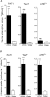
Figure 1
Figure 1. β amino acid uptake in PAT1-, TauT- or ATB0,+-expressing oocytes
Uptake of [3H]taurine (A) and [3H]β-alanine (B) into oocytes injected with PAT1, TauT or ATB0,+ cRNA (filled bars). For PAT1, β-alanine and taurine (10 μm) uptakes were measured at pH 5.5 in the absence of extracellular Na+. For TauT and ATB0,+, β-alanine and taurine (2 μm), uptakes were measured at pH 7.4 in the presence of extracellular Na+. As a control, uptake into water-injected oocytes (open bars) was measured under identical conditions in all cases. Data are mean ±s.e.m. (n= 15–30). NS, P > 0.05; ***, P < 0.001; all versus water-injected oocytes.
Figure 2
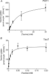
Figure 2. Concentration-dependent taurine uptake in PAT1- or TauT-expressing oocytes
Concentration-dependent [3H]taurine uptake was measured into oocytes injected with cRNA for PAT1 (A) and TauT (B). Uptake into PAT1-expressing oocytes was measured in Na+-free solutions at extracellular pH 5.5. Uptake into TauT-expressing oocytes was measured in NaCl-containing solutions at extracellular pH 7.4. Under each condition, uptake in water-injected oocytes was subtracted from that in cRNA-injected oocytes to give either PAT1- or TauT-specific uptake. Michaelis–Menten kinetics are fitted to the data which are mean ±s.e.m. (n= 7–10).
Figure 3
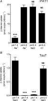
Figure 3. Distinct ion dependency of β amino acid uptake in PAT1- and TauT-expressing oocytes
A, [3H]β-alanine (10 μm) uptake was measured into PAT1 cRNA-injected oocytes at pH 5.5 in the presence of NaCl or in Na+-free (–Na+) or Cl-free (–Cl) conditions, and at pH 7.4 under Na+-free (–Na+) conditions. Uptake in water-injected oocytes was subtracted from that in cRNA-injected oocytes to give PAT1-specific uptake. Data are mean ±s.e.m. (n= 17–18). NS, P > 0.05; ***, P < 0.001; all versus pH 5.5 –Na+. B,[3H]β-alanine (2 μm) uptake was measured into TauT cRNA-injected oocytes at pH 7.4 in the presence of NaCl or in Na+-free (–Na+) or Cl-free (–Cl) conditions, or at pH 5.5 in the presence of NaCl. Uptake in water-injected oocytes was subtracted from that in cRNA-injected oocytes to give TauT-specific uptake. Data are mean ±s.e.m. (n= 9–10). NS, P > 0.05; ***, P < 0.001; all versus pH 7.4 NaCl.
Figure 4
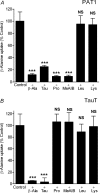
Figure 4. Inhibition of PAT1- or TauT-mediated β amino acid uptake by unlabelled amino acids
A, the ability of various amino acids and the amino acid analogue MeAIB (all 10 mm) to inhibit [3H]β-alanine (10 μm) uptake into PAT1-expressing oocytes was measured in Na+-free pH 5.5 solutions. Uptake in water-injected oocytes was subtracted from that in cRNA-injected oocytes to give PAT1-specific uptake. Data are mean ±s.e.m. (n= 19–20). NS, P > 0.05; ***, P < 0.001; all versus Control. B, the ability of various amino acids and the amino acid analogue MeAIB (all 500 μm) to inhibit [3H]β-alanine (2 μm) uptake into TauT-expressing oocytes was measured in NaCl-containing pH 7.4 solutions. Uptake in water-injected oocytes was subtracted from that in cRNA-injected oocytes to give TauT-specific uptake. Data are mean ±s.e.m. (n= 16–20). NS, P > 0.05; ***, P < 0.001; all versus Control.
Figure 5
Figure 5. Functional characteristics of taurine uptake across the brush-border membrane of human intestinal (Caco-2) cell monolayers demonstrate the involvement of both PAT1 and TauT
A, to demonstrate H+-coupled, pH-dependent, Na+-independent, PAT1-mediated transport, [3H]taurine uptake across the apical membrane of Caco-2 cell monolayers was measured at an extracellular taurine concentration of 100 μm under Na+-free conditions at apical pH 5.5 and 7.4 (basolateral pH 7.4). At pH 5.5, [3H]taurine uptake was measured in the presence and absence of the PAT1 substrates β-alanine and proline (both 10 mm). Data are mean ±s.e.m. (n= 10). ***, P < 0.001; all versus pH 5.5. B, concentration-dependent PAT1-mediated [3H]taurine uptake (0.1–30 mm taurine) was measured at apical pH 5.5 in the absence of extracellular Na+ (TauT will be inactive under these conditions). Michaelis–Menten kinetics are fitted to the data which are mean ±s.e.m. (n= 6). Inset, Eadie–Hofstee plot: V, taurine uptake; S, taurine concentration. C, to demonstrate Na+- and Cl-dependent TauT-mediated transport, [3H]taurine uptake across the apical membrane of Caco-2 cell monolayers was measured at an extracellular concentration of 0.1 μm taurine at apical pH 7.4 in NaCl-containing solutions and in the absence of extracellular Na+ (–Na+) or Cl (–Cl) (basolateral pH 7.4). In the presence of both Na+ and Cl, [3H]taurine uptake was measured in the presence and absence of the TauT substrate β-alanine (500 μm) or proline (500 μm) which is excluded from TauT at this concentration. Data are mean ±s.e.m. (n= 10). NS, P > 0.05; ***, P < 0.001; all versus NaCl. D, concentration-dependent TauT-mediated [3H]taurine uptake (0.0001–0.05 mm taurine) was measured at apical pH 7.4 in NaCl-containing solutions. Michaelis–Menten kinetics are fitted to the data which are mean ±s.e.m. (n= 6). Inset, Eadie–Hofstee plot (filled symbols represent data in main panel D over taurine concentration range 0.0001–0.05 mm; open symbols represent data for uptake at 0.1 and 0.2 mm): V, taurine uptake; S, taurine concentration.
Figure 6
Figure 6. The relative contributions of PAT1 and TauT to total taurine uptake across the brush-border membrane of human intestinal (Caco-2) cell monolayers varies with the luminal taurine concentration
Taurine (0.0001–1 mm) uptake across the apical membrane of Caco-2 cell monolayers was measured at the physiological apical pH of 6.5 (basolateral pH 7.4) in the presence and absence of extracellular Cl. Removal of extracellular Cl will selectively inhibit TauT whereas PAT1 will remain active. At each extracellular taurine concentration, taurine uptake is expressed as a percentage of the total uptake in the presence of Cl. The PAT1-mediated component is taken as the uptake in the absence of Cl (filled columns) and the TauT-mediated component is the difference between uptake in the presence and absence of Cl (open columns). Data are mean ±s.e.m. (n= 10).
Figure 7
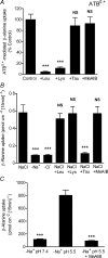
Figure 7. ATB0,+ has a distinct substrate selectivity, compared to PAT1 and TauT, and does not contribute to β-alanine uptake across the brush-border membrane of human intestinal (Caco-2) cell monolayers
A, [3H]β-alanine (100 μm) uptake into ATB0,+-expressing oocytes was measured in NaCl-containing, pH 7.4 solutions in the presence and absence of various amino acids and the amino acid analogue MeAIB (all 2 mm). Uptake in water-injected oocytes was subtracted from that in cRNA-injected oocytes to give ATB0,+-specific uptake. Data are mean ±s.e.m. (n= 16–20). NS, P > 0.05; ***, P < 0.001; all versus Control. B: [3H]β-alanine uptake across the brush-border membrane of Caco-2 cell monolayers was measured under conditions favourable for both TauT and ATB0,+ function (0.1 μmβ-alanine, pH 7.4 NaCl-containing apical solution). [3H]β-Alanine uptake was measured in the presence (NaCl) or absence of either Na+ (–Na+) or Cl (–Cl). [3H]β-Alanine uptake was also measured at pH 7.4 in the presence of NaCl and in the presence and absence of various amino acids at concentrations that discriminate between function of ATB0,+, TauT and PAT1: 2.5 mm leucine (to inhibit ATB0,+); 2.5 mm lysine (to inhibit ATB0,+); 500 μm taurine (to inhibit TauT); 30 mm MeAIB (to inhibit PAT1). Data are mean ±s.e.m. (n= 12). NS, P > 0.05; ***, P < 0.001; all versus NaCl. C, pH-dependent PAT1-mediated β-alanine uptake across the apical membrane of Caco-2 cell monolayers is observed when [3H]β-alanine uptake is measured under conditions favourable for PAT1 function (100 μmβ-alanine, apical pH 5.5, Na+-free solutions). Inhibition of [3H]β-alanine uptake was observed in the presence of the PAT1 substrate MeAIB (30 mm). Data are mean ±s.e.m. (n= 11–12). ***, P < 0.001; versus–Na+ pH 5.5.
Figure 8
Figure 8. Real-time PCR detection of PAT1, TauT and ATB0,+ transcripts in a human gastrointestinal tract cDNA panel and human mucosal biopsy samples
Expression of PAT1, TauT and ATB0,+ transcripts in a human gastrointestinal tract cDNA panel (A–C) and human mucosal biopsy samples (D–F). The tissues investigated were stomach (Stom), duodenum (Duo), jejunum (Jej), ileum (Ile), ascending colon (Asc), transverse colon (Tran) and descending colon (Des). The cDNA panel samples each comprised a pool of cDNAs from multiple donors and were assayed in duplicate whereas human duodenal mucosal biopsy samples were n= 4–5 (individual patients) and ileal samples n= 8 (individual patients). Data are expressed as mRNA abundance relative to GAPDH (pg/ng GAPDH). NS, P > 0.05; **, P < 0.01; all versus duodenum.

Similar articles

Cited by

References

    1. Anderson CMH, Ganapathy V, Thwaites DT. High- and low-capacity taurine transport across the luminal membrane of the small intestinal epithelium. FASEB J. 2008a;22:1202.3.
    1. Anderson CMH, Ganapathy V, Thwaites DT. Human solute carrier SLC6A14 is the β-alanine carrier. J Physiol. 2008b;586:4061–4067. - PMC - PubMed
    1. Anderson CMH, Grenade DS, Boll M, Foltz M, Wake KA, Kennedy DJ, Munck LK, Miyauchi S, Taylor PM, Campbell FC, Munck BG, Daniel H, Ganapathy V, Thwaites DT. H+/amino acid transporter 1 (PAT1) is the imino acid carrier: an intestinal nutrient/drug transporter in human and rat. Gastroenterology. 2004;127:1410–1422. - PubMed
    1. Anderson CMH, Thwaites DT. Indirect regulation of the intestinal H+-coupled amino acid transporter hPAT1 (SLC36A1) J Cell Physiol. 2005;204:604–613. - PubMed
    1. Balesaria S, Pell RJ, Abbott LJ, Tasleem A, Chavele KM, Barley NF, Khair U, Simon A, Moriarty KJ, Brydon WG, Walters JRF. Exploring possible mechanisms for primary bile acid malabsorption: evidence for different regulation of ileal bile acid transporter transcripts in chronic diarrhoea. Eur J Gastroenterol Hepatol. 2008;20:413–422. - PubMed

Publication types