

Reduced adipose tissue oxygenation in human obesity: evidence for rarefaction, macrophage chemotaxis, and inflammation without an angiogenic response
- PMID: 19074987
- PMCID: PMC2646071
- DOI: 10.2337/db08-1098
Reduced adipose tissue oxygenation in human obesity: evidence for rarefaction, macrophage chemotaxis, and inflammation without an angiogenic response
Abstract
Objective: Based on rodent studies, we examined the hypothesis that increased adipose tissue (AT) mass in obesity without an adequate support of vascularization might lead to hypoxia, macrophage infiltration, and inflammation.
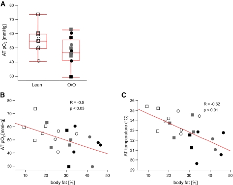
Research design and methods: Oxygen partial pressure (AT pO2) and AT temperature in abdominal AT (9 lean and 12 overweight/obese men and women) was measured by direct insertion of a polarographic Clark electrode. Body composition was measured by dual-energy X-ray absorptiometry, and insulin sensitivity was measured by hyperinsulinemic-euglycemic clamp. Abdominal subcutaneous tissue was used for staining, quantitative RT-PCR, and chemokine secretion assay.
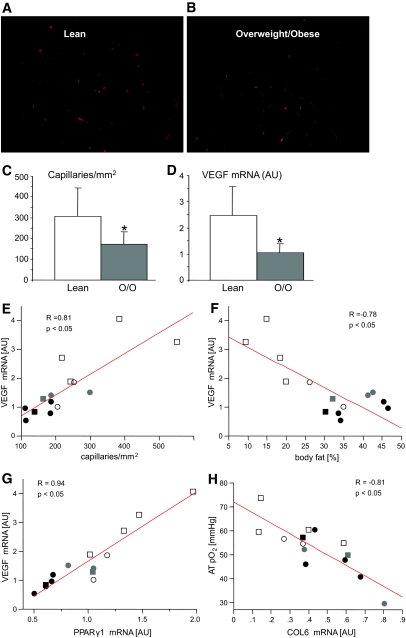
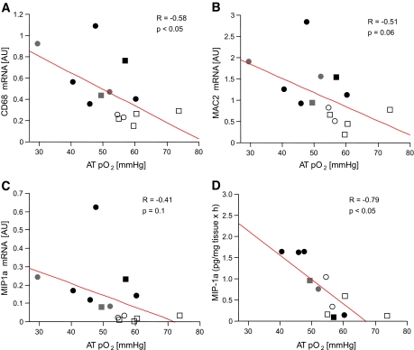
Results: AT pO2 was lower in overweight/obese subjects than lean subjects (47 +/- 10.6 vs. 55 +/- 9.1 mmHg); however, this level of pO2 did not activate the classic hypoxia targets (pyruvate dehydrogenase kinase and vascular endothelial growth factor [VEGF]). AT pO2 was negatively correlated with percent body fat (R = -0.50, P < 0.05). Compared with lean subjects, overweight/obese subjects had 44% lower capillary density and 58% lower VEGF, suggesting AT rarefaction (capillary drop out). This might be due to lower peroxisome proliferator-activated receptor gamma1 and higher collagen VI mRNA expression, which correlated with AT pO2 (P < 0.05). Of clinical importance, AT pO2 negatively correlated with CD68 mRNA and macrophage inflammatory protein 1alpha secretion (R = -0.58, R = -0.79, P < 0.05), suggesting that lower AT pO2 could drive AT inflammation in obesity.
Conclusions: Adipose tissue rarefaction might lie upstream of both low AT pO2 and inflammation in obesity. These results suggest novel approaches to treat the dysfunctional AT found in obesity.
Figures
References
-
- Smith SR, Ravussin E: Role of Adipocyte in Metabolism and Endocrine Function. DeGroot, Jameson, Endocrinology, Philadelphia, Elsevier Saunders, 2006
-
- Danforth E Jr: Failure of adipocyte differentiation causes type II diabetes mellitus? Nat Genet 26: 13, 2000 - PubMed
-
- Nishimura S, Manabe I, Nagasaki M, Hosoya Y, Yamashita H, Fujita H, Ohsugi M, Tobe K, Kadowaki T, Nagai R, Sugiura S: Adipogenesis in obesity requires close interplay between differentiating adipocytes, stromal cells, and blood vessels. Diabetes 56: 1517–1526, 2007 - PubMed
-
- Hausman GJ, Richardson RL: Adipose tissue angiogenesis. J Anim Sci 82: 925–934, 2004 - PubMed
-
- Lijnen HR, Christiaens V, Scroyen I, Voros G, Tjwa M, Carmeliet P, Collen D: Impaired adipose tissue development in mice with inactivation of placental growth factor function. Diabetes 55: 2698–2704, 2006 - PubMed
