. 2009 Feb;29(4):1059-71.
doi: 10.1128/MCB.01062-08. Epub 2008 Dec 15.

Aurora-A kinase is essential for bipolar spindle formation and early development

Affiliations

Aurora-A kinase is essential for bipolar spindle formation and early development

Dale O Cowley et al. Mol Cell Biol. 2009 Feb.

Abstract

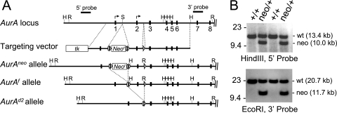
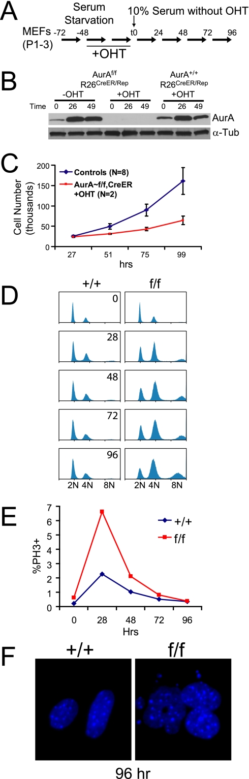
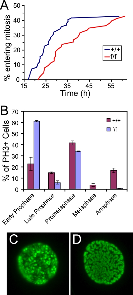
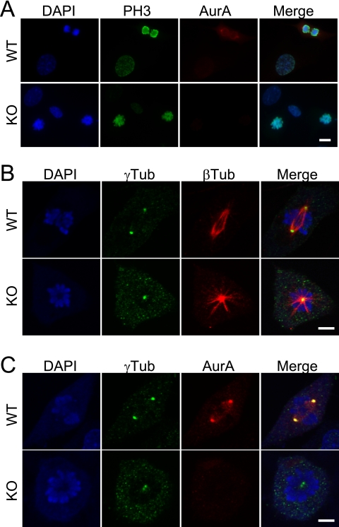
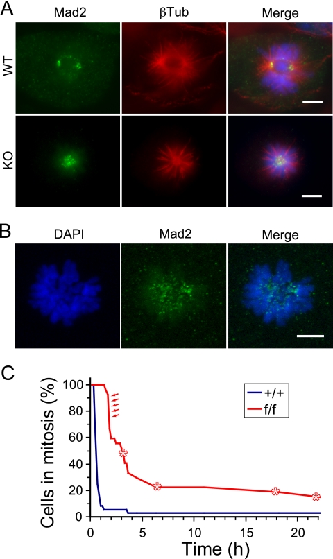
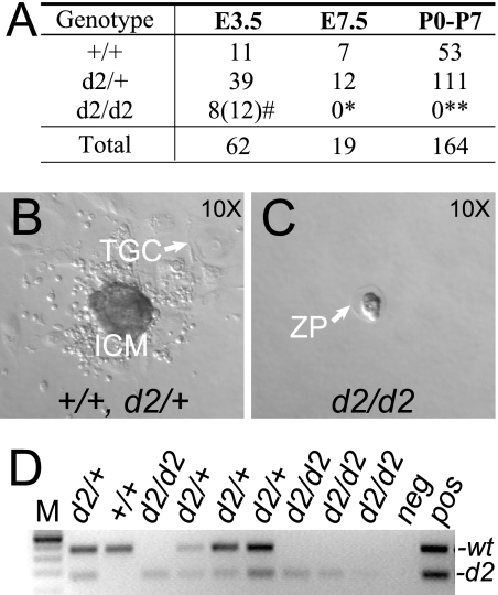
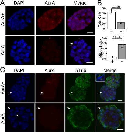
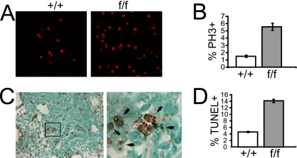
Aurora-A is a conserved kinase implicated in mitotic regulation and carcinogenesis. Aurora-A was previously implicated in mitotic entry and spindle assembly, although contradictory results prevented a clear understanding of the roles of Aurora-A in mammals. We developed a conditional null mutation in the mouse Aurora-A gene to investigate Aurora-A functions in primary cells ex vivo and in vivo. We show here that conditional Aurora-A ablation in cultured embryonic fibroblasts causes impaired mitotic entry and mitotic arrest with a profound defect in bipolar spindle formation. Germ line Aurora-A deficiency causes embryonic death at the blastocyst stage with pronounced cell proliferation failure, mitotic arrest, and monopolar spindle formation. Aurora-A deletion in mid-gestation embryos causes an increase in mitotic and apoptotic cells. These results indicate that murine Aurora-A facilitates, but is not absolutely required for, mitotic entry in murine embryonic fibroblasts and is essential for centrosome separation and bipolar spindle formation in vitro and in vivo. Aurora-A deletion increases apoptosis, suggesting that molecular therapies targeting Aurora-A may be effective in inducing tumor cell apoptosis. Aurora-A conditional mutant mice provide a valuable system for further defining Aurora-A functions and for predicting effects of Aurora-A therapeutic intervention.


Figures

FIG. 1.
FIG. 1.
Production of AurA conditional and mutant alleles. (A) Schematic representation of the AurA locus, targeting vector, and mutant alleles. Exons 1 to 8; Southern blot probes; and HindIII (H), EcoRI (R), and SalI (S) restriction sites are shown. Arrows indicate potential translational start codons. The targeting vector included a thymidine kinase gene (tk) and a neomycin resistance gene (Neor) flanked by FRT (ovals) and loxP (triangles) sites. Mice carrying the AurAneo allele were crossed to a Cre recombinase strain to generate the AurAf (conditional) and AurAd2 (null) alleles. (B) Southern blotting of ES cells indicating correct targeting event in the second and fourth clones from the left.
FIG. 2.
FIG. 2.
AurA mutation causes MEF growth defects and ploidy changes. (A) Experimental protocol for AurA deletion in quiescent MEFs. Early-passage MEFs were serum starved for 24 h and then serum starved for 48 h with OHT. The time of serum addition (without OHT) is defined as time zero. (B) Immunoblot analysis of AurA protein expression in OHT-treated MEFs shows that AurA protein is undetectable following Cre recombination. Cells were treated as shown in panel A, and samples were harvested at 0, 26, and 49 h after serum addition. α-Tubulin (α-Tub) detection confirmed equal loading. (C) MEF growth curve following the treatment protocol from panel A. Controls were AurAf/f; R26CreER/Rep cells with OHT treatment (n = 2), AurA+/+; R26CreER/Rep cells with or without OHT (n = 2 each), and AurAf/f; R26Rep/Rep cells with or without OHT (n = 1 each). (D) AurA deletion causes increased G2/M and endoreduplicating fractions. Cultures were treated with OHT as in panel A and analyzed by fluorescence-activated cell sorting for DNA content with propidium iodide. +/+ = AurA+/+; R26CreER/Rep; f/f = AurAf/f; R26CreER/Rep. (E) AurA deletion causes an increased PH3+ index. Cultures were treated with OHT as in panel A and analyzed by fluorescence-activated cell sorting for PH3 staining. +/+ = AurA+/+; R26CreER/Rep; f/f = AurAf/f; R26CreER/Rep. (F) Nuclear atypia following AurA deletion. DAPI-stained images from cultures 96 h after serum readdition are shown.
FIG. 3.
FIG. 3.
AurA-deficient cells have a mitotic entry defect. (A). Line graphs of time-lapse imaging data showing that AurA mutant cultures are delayed in mitotic entry after serum starvation. Graphs indicate the cumulative percentage of cells that entered mitosis from 17 to 61 h after serum addition. The experiment was repeated twice, with total numbers of 84 AurA+/+; R26CreER/+ (+/+) and 63 AurAf/f; R26CreER/+ (f/f) samples. (B). Bar graph showing the percentage of PH3+ cells in each mitotic phase 48 h after serum addition. Cells on coverslips were synchronized, treated with OHT, and released as described in the legend to Fig. 2A. Coverslips were fixed 48 h after serum addition, stained with anti-PH3 antibody, and manually scored for mitotic phase. Genotypes are as described in the legend to Fig. 2E. The experiment was performed with MEFs from two different embryos per genotype, and 200 random PH3+ cells were scored for each MEF line. t tests were used to compare percentages in each phase between genotypes: early prophase (P = 0.0108), late prophase (P = 0.0295), prometaphase (P = 0.0422), metaphase (P = 0.0572), and anaphase (P = 00092). (C). Example of PH3 antibody staining in an early prophase cell. (D). Example of PH3 antibody staining in a late prophase cell.
FIG. 4.
FIG. 4.
AurA-deficient cells that enter mitosis form monopolar spindles. (A) AurA-deficient cells enter mitosis and phosphorylate histone H3. DAPI, PH3, and AurA staining in WT (AurA+/+; R26CreER/Rep +OHT) and AurA-deficient (AurAf/f; R26CreER/Rep +OHT) MEFs 24 h after serum addition. Note the PH3-positive mitotic cells lacking AurA staining in the knockout (KO) part of the panel. Scale bar = 10 μm. (B) AurA-deficient cells form monopolar spindles. Confocal images of WT and KO cells stained for DAPI, γ-tubulin (γTub; centrosomes), and β-tubulin (βTub; spindles) are shown. The WT cell shows a normal bipolar prometaphase configuration. Note the characteristic monopolar spindle in the KO cell, including the circular chromosome array surrounding the single γ-tubulin focus with an astral microtubule array. Scale bar = 5 μm. (C) Centrosomes duplicate but fail to separate in AurA-deficient cells. Confocal images of WT and KO cells stained for DAPI, γ-tubulin (centrosomes), and AurA are shown. AurA is localized on centrosomes and adjacent spindle structures of the WT cell. Note the two closely associated γ-tubulin foci in the KO cell. Scale bar = 5 μm.
FIG. 5.
FIG. 5.
Monopolar spindles in AurA-deficient cells activate the spindle checkpoint. (A) Mad2 stains kinetochores of AurA-deficient cells. Cells were stained for Mad2, β-tubulin (βTub), and DAPI (shown as a merged image). WT cell shows Mad2 foci at poles and at unattached kinetochores. Multiple Mad2 foci are visible in the central region of the monopolar spindle in the AurA mutant cell, consistent with kinetochore localization (bottom). Scale bars = 5 μm. (B) Confocal maximum projection image of DAPI and Mad2 staining in an AurA mutant cell. Note the Mad2 foci in the central region near the ends of chromosomes where kinetochores are located. Scale bar = 5 μm. (C) AurA mutant cells experience mitotic delays and fail to divide. Line graph of time-lapse imaging data depicting times spent in mitosis by AurA+/+; R26CreER/+ (+/+) and AurAf/f; R26CreER/+ (f/f) cells. Asterisks indicate cells that died. Red arrows indicate f/f cells that divided. All other f/f cells died or exited mitosis without dividing, while 35/36 +/+ cells divided (see Table S2 and Fig. S2 in the supplemental material).
FIG. 6.
FIG. 6.
AurAd2/d2 embryos die at the blastocyst stage. (A) Genotype analysis of progeny from AurAd2/+ intercrosses at E3.5, E7.5, and postnatal stages. E3.5 embryos were cultured to generate blastocyst outgrowths and then genotyped. #, 12 embryos displayed similar in vitro phenotypes, but genotyping was only successful for 8; P > 0.05 by χ2 test for Mendelian segregation (including 12 presumed d2/d2 embryos). *, E7.5, P < 0.05. **, postnatal days 0 to 7 (P0-P7), P < 0.0001. (B) In vitro blastocyst outgrowth characteristic of the AurA+/+ and AurAd2/+ genotypes. ICM, cell clump derived from inner cell mass; TGC, trophoblast-derived giant cell. (C) AurAd2/d2 blastocyst after 7 days in culture. AurA mutant blastocysts remained unhatched within the zona pellucida (ZP). (D) Representative PCR genotyping results for blastocysts after in vitro culture. Deduced genotypes are shown at the top. Positions of WT and d2 products are indicated on the right. M, DNA ladder; neg, reactions run without template DNA; pos, positive control reactions.
FIG. 7.
FIG. 7.
Mitotic arrest and monopolar spindles in AurAd2/d2 blastocysts. (A) Confocal maximum z projection of E3.5 blastocysts after whole-mount IF staining. Top: AurA+/+ or AurAd2/+ embryo with a clear AurA signal at the centrosomes of mitotic cells. DAPI was used to stain DNA. Mitotic cells are identifiable by condensed chromosomes. Bottom: AurAd2/d2 embryo. Note the lack of AurA staining and the presence of multiple mitotic cells with condensed chromatin. The arrow indicates a circular chromosome array indicative of a monopolar spindle. Scale bars = 20 μm. (B) AurAd2/d2 blastocysts have significantly reduced cell numbers (top) and an increased mitotic index (bottom) relative to those of WT littermates. Embryos stained as described for panel A were scored as WT (AurA+/+ or AurAd2/+) or AurAd2/d2 mutants based on the presence or absence of AurA staining. Cells were counted in serial confocal z sections and scored as mitotic or nonmitotic on the basis of chromatin condensation. Histograms show mean values for three embryos per class. Error bars indicate standard errors. P values were derived from two-tailed t tests. (C) Apoptosis and monopolar spindles in AurAd2/d2 embryos. Top: AurA+/+ or AurAd2/+ embryo with normal α-tubulin (αTub) staining. The arrow indicates a metaphase cell with a bipolar spindle. Bottom: AurAd2/d2 embryo. Note the multiple apoptotic bodies (arrowhead) and cells with circular chromosome arrays and astral microtubule patterns indicative of a monopolar spindle phenotype (arrow). Images are confocal maximum z projections of a subset of z slices for each embryo. Scale bars = 20 μm.
FIG. 8.
FIG. 8.
AurA deletion in mid-gestation embryos causes an increased mitotic index and apoptosis. (A). Representative PH3 staining in lung sections from E14.5 embryos after tamoxifen treatment at E12.5. (B). Quantitation of PH3+ cells in lung tissue from E14.5 embryos after tamoxifen treatment at E12.5. (C). Left: representative TUNEL of a lung section from an E14.6 AurAf/f; R26CreER/Rep embryo after tamoxifen treatment at E12.5. Right: magnified image from the box in the left part of the panel showing examples of TUNEL+ cells (brown). (D). Quantitation of TUNEL+ cells in lung tissue from an E14.5 embryo after tamoxifen treatment at E12.5.

References

    1. Andrews, P. D., E. Knatko, W. J. Moore, and J. R. Swedlow. 2003. Mitotic mechanics: the auroras come into view. Curr. Opin. Cell Biol. 15672-683. - PubMed
    1. Badea, T. C., Y. Wang, and J. Nathans. 2003. A noninvasive genetic/pharmacologic strategy for visualizing cell morphology and clonal relationships in the mouse. J. Neurosci. 232314-2322. - PMC - PubMed
    1. Berdnik, D., and J. A. Knoblich. 2002. Drosophila Aurora-A is required for centrosome maturation and actin-dependent asymmetric protein localization during mitosis. Curr. Biol. 12640-647. - PubMed
    1. Bergnes, G., K. Brejc, and L. Belmont. 2005. Mitotic kinesins: prospects for antimitotic drug discovery. Curr. Top. Med. Chem. 5127-145. - PubMed
    1. Bischoff, J. R., L. Anderson, Y. Zhu, K. Mossie, L. Ng, B. Souza, B. Schryver, P. Flanagan, F. Clairvoyant, C. Ginther, C. S. Chan, M. Novotny, D. J. Slamon, and G. D. Plowman. 1998. A homologue of Drosophila aurora kinase is oncogenic and amplified in human colorectal cancers. EMBO J. 173052-3065. - PMC - PubMed

Publication types

MeSH terms