

Deficiency of the tetraspanin CD63 associated with kidney pathology but normal lysosomal function
- PMID: 19075008
- PMCID: PMC2643809
- DOI: 10.1128/MCB.01163-08
Deficiency of the tetraspanin CD63 associated with kidney pathology but normal lysosomal function
Abstract
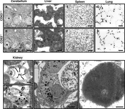
CD63 is a member of the tetraspanin superfamily that constitutes a main component of the lysosomal membrane. In mice, two CD63 gene loci are present, with only one of these two being functional. We generated and analyzed mice deficient for active CD63. Disruption of CD63 results in a complete loss of CD63 protein expression. Despite its abundance in late endosomes/lysosomes, the lack of CD63 does not cause obvious endosomal/lysosomal abnormalities. CD63 knockout mice are viable and fertile without gross morphological abnormalities in the majority of tissues. No alterations in the populations of immune cells and only minor differences in platelet function were observed. This suggests that the lack of CD63 could be successfully compensated for, most likely by other tetraspanins. However, CD63 deficiency leads to an altered water balance. CD63 knockout mice show an increased urinary flow, water intake, reduced urine osmolality, and a higher fecal water content. In principle cells of the collecting duct of CD63-deficient mice, abnormal intracellular lamellar inclusions were observed. This indicates that the sorting of apical transport proteins might be impaired in these cells. CD63 knockout mice provide an important tool for analyzing the various postulated functions of CD63 in vivo.
Figures


Similar articles
-
Role of adaptor complex AP-3 in targeting wild-type and mutated CD63 to lysosomes.Mol Biol Cell. 2002 Mar;13(3):1071-82. doi: 10.1091/mbc.01-08-0409. Mol Biol Cell. 2002. PMID: 11907283 Free PMC article.
-
CD63 antigen. A novel lysosomal membrane glycoprotein, cloned by a screening procedure for intracellular antigens in eukaryotic cells.J Biol Chem. 1991 Feb 15;266(5):3239-45. J Biol Chem. 1991. PMID: 1993697
-
CD63 tetraspanin slows down cell migration and translocates to the endosomal-lysosomal-MIICs route after extracellular stimuli in human immature dendritic cells.Blood. 2004 Aug 15;104(4):1183-90. doi: 10.1182/blood-2004-01-0104. Epub 2004 May 6. Blood. 2004. PMID: 15130945
-
Trafficking and function of the tetraspanin CD63.Exp Cell Res. 2009 May 15;315(9):1584-92. doi: 10.1016/j.yexcr.2008.09.020. Epub 2008 Oct 7. Exp Cell Res. 2009. PMID: 18930046 Review.
-
Intracellular trafficking of lysosomal membrane proteins.Bioessays. 1996 May;18(5):379-89. doi: 10.1002/bies.950180508. Bioessays. 1996. PMID: 8639161 Review.
Cited by
-
Adaptor protein complex 2-mediated, clathrin-dependent endocytosis, and related gene activities, are a prominent feature during maturation stage amelogenesis.J Bone Miner Res. 2013 Mar;28(3):672-87. doi: 10.1002/jbmr.1779. J Bone Miner Res. 2013. PMID: 23044750 Free PMC article.
-
Cell biology and physiology of the uroepithelium.Am J Physiol Renal Physiol. 2009 Dec;297(6):F1477-501. doi: 10.1152/ajprenal.00327.2009. Epub 2009 Jul 8. Am J Physiol Renal Physiol. 2009. PMID: 19587142 Free PMC article. Review.
-
Platelet integrin αIIbβ3: signal transduction, regulation, and its therapeutic targeting.J Hematol Oncol. 2019 Mar 7;12(1):26. doi: 10.1186/s13045-019-0709-6. J Hematol Oncol. 2019. PMID: 30845955 Free PMC article. Review.
-
Generation of a novel transgenic rat model for tracing extracellular vesicles in body fluids.Sci Rep. 2016 Aug 19;6:31172. doi: 10.1038/srep31172. Sci Rep. 2016. PMID: 27539050 Free PMC article.
-
Rapid Isolation of Rare Isotype-Switched Hybridoma Variants: Application to the Generation of IgG2a and IgG2b MAb to CD63, a Late Endosome and Exosome Marker.Antibodies (Basel). 2020 Jul 2;9(3):29. doi: 10.3390/antib9030029. Antibodies (Basel). 2020. PMID: 32630723 Free PMC article.
References
-
- Atkinson, B., C. S. Ernst, B. F. D. Ghrist, M. Herlyn, M. Blaszczyk, A. H. Ross, D. Herlyn, Z. Steplewski, and H. Koprowski. 1984. Identification of melanoma-associated antigens using fixed tissue screening of antibodies. Cancer Res. 442577-2581. - PubMed
-
- Atkinson, B., C. S. Ernst, B. F. D. Ghrist, A. H. Ross, W. H. Clark, M. Herlyn, D. Herlyn, G. Maul, Z. Steplewski, and H. Koprowski. 1985. Monoclonal antibody to a highly glycosylated protein reacts in fixed tissue with melanoma and other tumors. Hybridoma 4243-255. - PubMed
-
- Azorsa, D. O., J. A. Hyman, and J. E. K. Hildreth. 1991. Cd63/Pltgp40: a platelet activation antigen identical to the stage-specific, melanoma-associated antigen Me491. Blood 78280-284. - PubMed
-
- Beinert, T., S. Munzing, K. Possinger, and F. Krombach. 2000. Increased expression of the tetraspanins CD53 and CD63 on apoptotic human neutrophils. J. Leukoc. Biol. 67369-373. - PubMed
Publication types
MeSH terms
Substances
LinkOut - more resources
Full Text Sources
Other Literature Sources
Medical
Molecular Biology Databases
Miscellaneous