. 2009 Feb 20;284(8):5343-51.
doi: 10.1074/jbc.M808430200. Epub 2008 Dec 15.

UAF1 is a subunit of multiple deubiquitinating enzyme complexes

Affiliations

UAF1 is a subunit of multiple deubiquitinating enzyme complexes

Martin A Cohn et al. J Biol Chem. .

Abstract

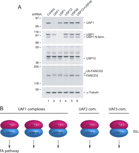
A balance between ubiquitination and deubiquitination regulates numerous cellular processes and pathways, and specific deubiquitinating enzymes often play the decisive role of controlling this balance. We recently reported that the USP1 deubiquitinating enzyme, which regulates the Fanconi anemia pathway by deubiquitinating the central player of the pathway, FANCD2, is activated by the WD40-repeat containing UAF1 protein through formation of a stable USP1/UAF1 protein complex. Here we present the isolation of two novel multisubunit deubiquitinating enzyme complexes containing USP12 and USP46, respectively. Both complexes contain the UAF1 protein as a bona fide subunit. Interestingly, UAF1 regulates the enzymatic activity of both enzyme complexes, suggesting that this activator protein may regulate a subclass of human deubiquitinating enzymes. We postulate that additional WD40-containing proteins may also form complexes with other human deubiquitinating enzymes and thereby regulate their activity and substrate specificity.


Figures

FIGURE 1.
FIGURE 1.
Purification of native UAF1 complexes containing USP1 and other deubiquitinating enzymes. A, Flag-HA-tagged UAF1 was immunoprecipitated from either HeLa cell extracts or cell extracts prepared from HeLa cells where the endogenous USP1 protein is stably knocked down by shRNA. The immunoprecipitate was analyzed by immunoblotting using the indicated antibodies. B, the immunoprecipitates from A were analyzed for associated deubiquitinating enzyme activity using ubiquitin-AMC as a substrate. C, silver stain of the UAF1 complexes. D, glycerol gradient sedimentation of the UAF1 complexes. Fractions were separated by SDS-PAGE, and proteins were visualized by silver stain. Subcomplexes as determined by sedimentation profile are indicated by roman numerals I-IV. Sedimentation profile of molecular weight markers is indicated. E, the fractions from C were analyzed by immunoblotting using the indicated antibodies (upper panels) and for deubiquitinating enzyme activity using ubiquitin-AMC (lower panel).
FIGURE 2.
FIGURE 2.
Identification of the USP12- and USP46-containing UAF1 complexes. A, fractions 3 and 5 fromtheglycerol-sedimented UAF1complex were incubated invitro with ubiquitin-VS and analyzed by silver stain. Arrows indicate USP1 and USP1-ubiquitin-VS complex (lanes 1-2). Likewise, arrows indicate putative deubiquitinating enzymes with molecular weights of 40 and 41 kDa, unbound or bound to ubiquitin-VS (lanes 3-4). In lane 5 only ubiquitin-VS is added. B, USP12 was identified by tandem mass spectrometry. The MS/MS spectrum of the peptide Leu-286-Arg-298 is shown. C, schematic of the USP12, UPS46 and USP1-deubiquitinating enzymes. Positions of the catalytic triad cysteine, histidine, and aspartic acid residues are indicated, as well as the position of the conserved asparagine. D, immunoblot analysis of the UAF1 complex using the indicated antibodies. E, silver stain of the USP12 complex. F, immunoblot analysis of the USP12 complex using the indicated antibodies.
FIGURE 3.
FIGURE 3.
UAF1 activates USP12 and USP46. A, Coomassie Blue stain of the proteins purified from Sf9 cells. B, in vitro enzymatic activity of USP12, USP12/UAF1, USP46, USP46/UAF1, USP1, and USP1/UAF1 was assayed using Ub-AMC as a substrate. The concentration of each protein was 100 nm except for USP1 and USP1/UAF1, which were 20 nm.
FIGURE 4.
FIGURE 4.
The WD40 repeats of UAF1 are required for the interaction with USP12 and USP46. A, scheme of wild-type UAF1 and deletion mutants thereof. B, Flag-tagged deletion mutants of UAF1 were transiently expressed in 293T cells. The Flag-tagged proteins were immunoprecipitated from cell lysates using anti-Flag antibodies. The immunoprecipitates were analyzed using the indicated antibodies.
FIGURE 5.
FIGURE 5.
UAF1 forms three distinct deubiquitinating enzyme complexes with USP1, USP12, and USP46. A, native UAF1, USP1, USP12, and USP46 complexes were purified from HeLa cells and analyzed by immunoblot analysis using the indicated antibodies. B, alignment of the amino acids surrounding the catalytic cysteine residue in USP12 and USP46. C, immunoblot analysis of the USP12 and USP12-C48S complexes using the indicated antibodies. D, USP12 and USP12-C48S complexes were analyzed for associated deubiquitinating enzyme activity using ubiquitin-AMC as a substrate.
FIGURE 6.
FIGURE 6.
USP12/UAF1 and USP46/UAF1 complexes do not regulate FANCD2 ubiquitination or the FA pathway. A, immunoblot analysis of protein lysates prepared from stable HeLa cell lines expressing shRNA constructs targeting UAF1, USP1, USP12, or USP46. The lysates were analyzed using the indicated antibodies. B, model for various deubiquitinating complexes. The USP1/UAF1, USP12/UAF1, and USP46/UAF1 complexes are shown schematically as indicated. The speculated protein complexes containing other human deubiquitinating enzymes and their activating subunits are shown on the right.

References

    1. Pickart, C. M., and Eddins, M. J. (2004) Biochim. Biophys. Acta 1695 55-72 - PubMed
    1. Kennedy, R. D., and D'Andrea, A. D. (2005) Genes Dev. 19 2925-2940 - PubMed
    1. Hoege, C., Pfander, B., Moldovan, G. L., Pyrowolakis, G., and Jentsch, S. (2002) Nature 419 135-141 - PubMed
    1. Sigismund, S., Polo, S., and Di Fiore, P. P. (2004) Curr. Top. Microbiol. Immunol. 286 149-185 - PubMed
    1. Hanna, J., and Finley, D. (2007) FEBS Lett. 581 2854-2861 - PMC - PubMed

Publication types

MeSH terms