. 2009 Mar;77(3):1022-30.
doi: 10.1128/IAI.01064-08. Epub 2008 Dec 15.

The emerging human pathogen Photorhabdus asymbiotica is a facultative intracellular bacterium and induces apoptosis of macrophage-like cells

Affiliations

The emerging human pathogen Photorhabdus asymbiotica is a facultative intracellular bacterium and induces apoptosis of macrophage-like cells

S C P Costa et al. Infect Immun. 2009 Mar.

Abstract

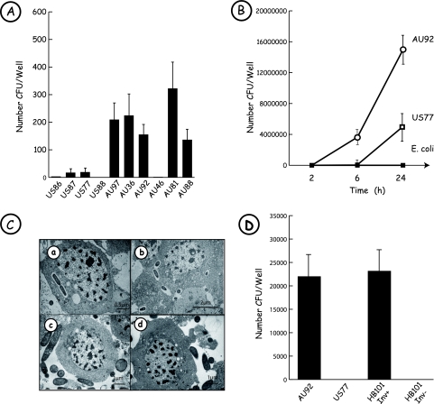
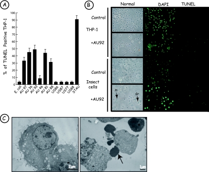
Photorhabdus species are gram-negative entomopathogenic bacteria of the family Enterobacteriaceae. Among the different members of the genus, one species, Photorhabdus asymbiotica, is a pathogen of both insects and humans. The pathogenicity mechanisms of this bacterium are unknown. Here we show that P. asymbiotica is a facultative intracellular pathogen that is able to replicate inside human macrophage-like cells. Furthermore, P. asymbiotica was shown for the first time in an intracellular location after insect infection. We also demonstrated that among Australian and American clinical isolates, only the Australian strains were able to invade nonphagocytic human cells. In cell culture infection experiments, Australian clinical isolates as well as cell-free bacterial culture supernatant induced strong apoptosis of a macrophage cell line at 6 h postinfection. American isolates also induced cellular death, but much later than that induced by Australian ones. Mammalian cultured cells analyzed for key features of apoptosis displayed apoptotic nuclear morphology, activation of the initiator caspases 8 and 9 and the executioner caspases 3 and 7, and poly(ADP-ribose) polymerase proteolysis, suggesting activation of both the intrinsic and extrinsic apoptotic pathways.


Figures

FIG. 1.
FIG. 1.
Photorhabdus asymbiotica is a facultative intracellular bacterium. (A) Comparison of Photorhabdus clinical strain uptake by human THP-1 cells. Infection experiments were performed at an MOI of 25:1 with PMA-differentiated THP-1 cells and with the different bacterial strains listed in Table 1. After 2 h of gentamicin treatment (100 μg/ml), macrophage-like cells were lysed, intracellular bacteria were recovered, and the number of CFU/well was determined. All Australian clinical isolates were engulfed by macrophages, except for strain AU46. The American isolates were weakly phagocytosed. Results are representative of three independent experiments. (B) Survival and growth of P. asymbiotica AU92 and US77 in human THP-1 cells. THP-1 cells were challenged with P. asymbiotica strains at an MOI of 25:1 and treated for 2 h with 100 μg/ml gentamicin. At different times postinfection, cells were lysed and bacteria plated. The American (open squares) and Australian (circles) clinical isolates studied were able to survive in THP-1 cells, in contrast to the nonpathogenic E. coli XL1-Blue strain (filled squares), which was eliminated by macrophage-like cells. Results are representative of three independent experiments. (C) Bacteria of clinical strains are present inside insect hemocytes. Hemocytes of M. unipuncta were incubated with P. asymbiotica AU92 (a and b), P. luminescens TT01 (c), or P. asymbiotica US77 (d). TEM observations show that the human clinical isolate P. asymbiotica AU92 is phagocytosed by hemocytes. In contrast, the strictly entomopathogenic organism P. luminescens, as well as P. asymbiotica US77, present an extracellular location. (D) Invasion of epithelial cells by P. asymbiotica. HeLa cells were infected for 1 h with the P. asymbiotica strains AU92 and US77 and with E. coli HB101 Inv+ (carrying the inv gene of Yersinia) and E. coli HB101 Inv as negative controls (MOI, 25:1). After 2 h of treatment with 100 μg/ml gentamicin, cells were lysed and intracellular bacteria plated. The Australian strain of P. asymbiotica was able to invade HeLa cells, with an intracellular uptake rate similar to that of E. coli HB101 harboring the Yersinia inv gene. Results are representative of three independent experiments.
FIG. 2.
FIG. 2.
Photorhabdus asymbiotica induces apoptosis of host cells. (A) Australian strains of P. asymbiotica induce apoptosis of THP-1 cells upon 6 h of infection. THP-1 cells were infected with the American and Australian Photorhabdus clinical strains at an MOI of 100:1 (E. coli XL1-Blue was used as a negative control, and staurosporine [STAU] was used as a positive control). TUNEL-positive cells were determined by fluorescence microscopy. At 6 h postinfection, all Australian strains induced apoptosis of THP-1 cells. Almost no TUNEL-positive THP-1 cells were found at 6 h postinfection when infection was done with the American strains. Results are representative of three independent experiments. (B) Apoptosis induced by P. asymbiotica strain AU92 is specific for macrophage-like cells. THP-1 cells and insect hemocytes were incubated for 6 h with P. asymbiotica at an MOI of 100:1. After DAPI staining, some THP-1 cells presented nuclear fragmentation, and these cells were TUNEL positive. Insect granulocytes (Gr) were TUNEL positive, in contrast to plasmatocytes (Pl). (C) Ultrastructure of THP-1 cells following infection with P. asymbiotica (by TEM). Control cells (left) and THP-1 cells (right) are shown after 6 h of incubation with AU92 (MOI, 100:1). Note the presence of typical apoptotic bodies (arrow).
FIG. 3.
FIG. 3.
Apoptosis induced by AU92 requires caspase activity. (A) Effects of various inhibitors on AU92-induced THP-1 cell apoptosis. THP-1 cells were incubated with 20 μM Z-VAD-FMK, 10 μM Z-IETD-FMK, 10 μM Z-LEHD-FMK, 100 nM wortmannin (Wort), or 5 μg/ml cytochalasin D (Cyto D) 20 min before infection. Cells were infected with P. asymbiotica AU92 at an MOI of 100 for 6 h, and TUNEL labeling was performed. The different caspase and phagocytosis inhibitors significantly reduced the number of TUNEL-positive cells after P. asymbiotica AU92 treatment. Staurosporine (2.5 μM) (Stau) was used as an apoptosis positive control. None, no treatment. Results are means ± SD for three independent experiments. *, P < 0.01. (B) P. asymbiotica AU92 and P. asymbiotica AU92 Sn activated caspases in THP-1 cells. THP-1 cells were incubated with P. asymbiotica AU92, AU92 Sn, US77, US77 Sn, or staurosporine. Caspase activities were determined by the released of luminescent products following the manufacturer's protocol. Caspase activities are expressed as percentages of those in staurosporine-treated cells. Results are means ± SD for three independent experiments. (C) P. asymbiotica induces caspase-3/7 and PARP cleavage. After 1 or 4 h of incubation of THP-1 cells with P. asymbiotica AU92 or bacterial supernatant (PaSn), total protein extracts were separated by 15% SDS-PAGE, followed by Western blotting with anti-caspase-3, anti-cleaved caspase-7, and anti-PARP antibodies. Native (around 35 kDa) and cleaved (around 20 kDa) forms of caspase-3, the cleaved form of caspase-7 (around 20 kDa), and PARP (around 85 kDa and 30 kDa) were already present after 1 h of incubation of THP-1 cells with PaSn. The same results were obtained with living but not with dead bacteria (data not shown). Staurosporine (2.5 μM) (Stau) was used as an apoptosis positive control. c, no treatment. Data are representative of three independent experiments.

References

    1. Akhurst, R. J., N. E. Boemare, P. H. Janssen, M. M. Peel, D. A. Alfredson, and C. E. Beard. 2004. Taxonomy of Australian clinical isolates of the genus Photorhabdus and proposal of Photorhabdus asymbiotica subsp. asymbiotica subsp. nov. and P. asymbiotica subsp. australis subsp. nov. Int. J. Syst. Evol. Microbiol. 541301-1310. - PubMed
    1. Bowen, D., T. A. Rocheleau, M. Blackburn, O. Andreev, E. Golubeva, R. Bhartia, and R. H. Ffrench-Constant. 1998. Insecticidal toxins from the bacterium Photorhabdus luminescens. Science 2802129-2132. - PubMed
    1. Brugirard-Ricaud, K., E. Duchaud, A. Givaudan, P. A. Girard, F. Kunst, N. Boemare, M. Brehélin, and R. Zumbihl. 2005. Site-specific antiphagocytic function of the Photorhabdus luminescens type III secretion system during insect colonization. Cell. Microbiol. 7363-371. - PubMed
    1. Brugirard-Ricaud, K., A. Givaudan, J. Parkhill, N. Boemare, F. Kunst, R. Zumbihl, and E. Duchaud. 2004. Variation in the effectors of the type III secretion system among Photorhabdus species as revealed by genomic analysis. J. Bacteriol. 1864376-4381. - PMC - PubMed
    1. Chowdhury, I., B. Tharakan, and G. K. Bhat. 2006. Current concepts in apoptosis: the physiological suicide program revisited. Cell. Mol. Biol. Lett. 11506-525. - PMC - PubMed

Publication types

MeSH terms

LinkOut - more resources