. 2009 Mar;77(3):1031-43.
doi: 10.1128/IAI.01192-08. Epub 2008 Dec 15.

Lactobacillus bulgaricus prevents intestinal epithelial cell injury caused by Enterobacter sakazakii-induced nitric oxide both in vitro and in the newborn rat model of necrotizing enterocolitis

Affiliations

Lactobacillus bulgaricus prevents intestinal epithelial cell injury caused by Enterobacter sakazakii-induced nitric oxide both in vitro and in the newborn rat model of necrotizing enterocolitis

Catherine J Hunter et al. Infect Immun. 2009 Mar.

Retraction in

Expression of concern in

Abstract

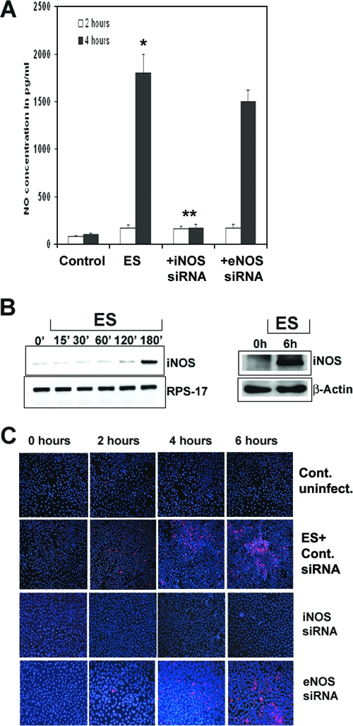
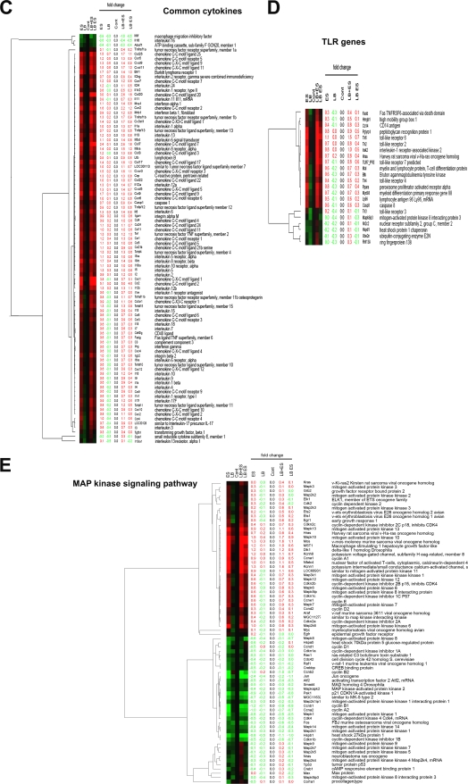
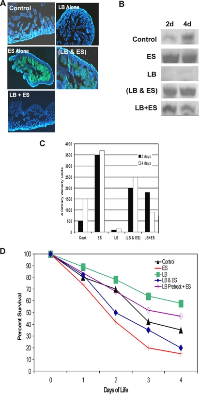
Enterobacter sakazakii is an emerging pathogen that has been associated with outbreaks of necrotizing enterocolitis (NEC) as well as infant sepsis and meningitis. Our previous studies demonstrated that E. sakazakii induces NEC in a newborn rat model by inducing enterocyte apoptosis. However, the mechanisms responsible for enterocyte apoptosis are not known. Here we demonstrate that E. sakazakii induces significant production of nitric oxide (NO) in rat intestinal epithelial cells (IEC-6) upon infection. The elevated production of NO, which is due to increased expression of inducible NO synthase, is responsible for apoptosis of IEC-6 cells. Notably, pretreatment of IEC-6 cells with Lactobacillus bulgaricus (ATCC 12278) attenuated the upregulation of NO production and thereby protected the cells from E. sakazakii-induced apoptosis. Furthermore, pretreatment with L. bulgaricus promoted the integrity of enterocytes both in vitro and in the infant rat model of NEC, even after challenge with E. sakazakii. Infection of IEC-6 cells with E. sakazakii upregulated several genes related to apoptosis, cytokine production, and various signaling pathways, as demonstrated by rat gene array analysis, and this upregulation was subdued by pretreatment with L. bulgaricus. In agreement with these data, L. bulgaricus pretreatment protected newborn rats infected with E. sakazakii from developing NEC, resulting in improved survival.


Figures

FIG. 1.
FIG. 1.
E. sakazakii-induced NO production is responsible for the apoptosis of IEC-6 cells. (A) Confluent monolayers of IEC-6 cells, either nontransfected or transfected with siRNA, were treated with E. sakazakii (ES) for 2 h or 4 h, the supernatants were collected and centrifuged to remove the bacteria, and NO production was determined by the Greiss method. Wild-type IEC-6 cells demonstrated a small increase in NO production by 2 h and a 10-fold increase after 6 h of exposure (*, P = 0.002), and NO production decreased significantly in siRNA-iNOS/IEC-6 cells (**, P < 0.001). The experiments were carried out separately in triplicate at least three times. (B) IEC-6 cells were infected with E. sakazakii for various periods, total RNA was extracted, and RT-PCR was performed using primers for iNOS and RPS-17. RNA expression was determined by semiquantitative PCR. In separate experiments, total cell lysates were prepared from IEC-6 cells infected with E. sakazakii for 6 h, and equal quantities of proteins were subjected to Western blotting using anti-iNOS or anti-β-actin antibody. (C) Confluent monolayers of IEC-6 cells, either nontransfected or transfected with siRNA to iNOS or eNOS, were left untreated (control) or infected with E. sakazakii for the indicated periods, and the monolayers were assessed for apoptosis by TUNEL staining using an ApoTag kit.
FIG. 2.
FIG. 2.
Pretreatment with L. bulgaricus prevents E. sakazakii-induced apoptosis in IEC-6 cells. (A) Total cell lysates of IEC-6 cells, either noninfected, infected with E. sakazakii (ES) or L. bulgaricus (LB), or pretreated with L. bulgaricus for 1 h followed by infection with E. sakazakii (LB+ES) were subjected to Western blotting with anti-iNOS or anti-β-actin antibody. A positive control for iNOS was also included in the blot. (B) Confluent monolayers of IEC-6 cells were infected with E. sakazakii for various periods and washed, and the number of bound E. sakazakii cells was determined as described in Materials and Methods. In separate experiments, IEC-6 cells were pretreated with L. bulgaricus for 1 h followed by E. sakazakii for the indicated periods. (C) IEC-6 cells infected with E. sakazakii or pretreated with L. bulgaricus for 1 h followed by E. sakazakii infection for 6 h were subjected to transmission electron microscopy. Magnification, ×6,500. (D) IEC-6 cells that were uninfected, infected with E. sakazakii, or pretreated with L. bulgaricus followed by E. sakazakii (LB+ES) for 6 h were subjected to TUNEL staining using an ApoTag kit. (E) IEC-6 cells were left uninfected (control), infected with GFP-labeled E. sakazakii alone (two panels of images are shown), or pretreated with L. bulgaricus for 1 followed by GFP-labeled E. sakazakii (LB+ES) for 4 h. The cells were stained with annexin V and 7-AAD as described in Materials and Methods and subjected to ImageStream analysis. The picture is representative of several cells screened for apoptosis.
FIG. 3.
FIG. 3.
L. bulgaricus pretreatment preserves the integrity of intestinal epithelial cells. (A) IEC-6 cells treated with bacteria as described in the legend to Fig. 2 and processed for scanning electron microscopy at 6 h postinfection. (B) Newborn rats were either left uninfected or infected with E. sakazakii by being fed 105 CFU once a day under hypoxic conditions. The animals were sacrificed after 4 days, intestines were collected, and sections of intestine were paraffin embedded and stained with hematoxylin and eosin. In separate experiments, newborn rats were either pretreated with 105 CFU of L. bulgaricus alone (c) or fed with L. bulgaricus and E. sakazakii together (d) or L. bulgaricus for 1 day followed by E. sakazakii (e). (C) The intestinal sections stained with hematoxylin and eosin were scored by a blinded pathologist and graphed. A protective effect of L. bulgaricus pretreatment is clearly seen in the presence of E. sakazakii compared with the effect of E. sakazakii alone (*, P < 0.001).
FIG. 4.
FIG. 4.
Gene expression analysis of IEC-6 cells infected with E. sakazakii in the presence or absence of L. bulgaricus. IEC-6 cells were either left uninfected, infected with E. sakazakii (ES), L. bulgaricus (LB), or L. bulgaricus and E. sakazakii together (LB+ES), or pretreated with L. bulgaricus for 1 h followed by E. sakazakii for 6 h (LB ES). Total RNA was isolated and used for whole rat genome DNA microarray analysis as described in Materials and Methods. Candidate genes for four categories were selected. (A) Apoptosis genes. (B) Unified pathway of apoptosis generated by KEGG indicating the upregulated genes in the rat genome or similar genes (red stars). (C) Common cytokine-related genes. (D) TLR genes. (E) MAPK signaling pathway genes. Genes exhibiting upregulation are represented in red, and downregulated genes are shown in green.
FIG. 4.
FIG. 4.
Gene expression analysis of IEC-6 cells infected with E. sakazakii in the presence or absence of L. bulgaricus. IEC-6 cells were either left uninfected, infected with E. sakazakii (ES), L. bulgaricus (LB), or L. bulgaricus and E. sakazakii together (LB+ES), or pretreated with L. bulgaricus for 1 h followed by E. sakazakii for 6 h (LB ES). Total RNA was isolated and used for whole rat genome DNA microarray analysis as described in Materials and Methods. Candidate genes for four categories were selected. (A) Apoptosis genes. (B) Unified pathway of apoptosis generated by KEGG indicating the upregulated genes in the rat genome or similar genes (red stars). (C) Common cytokine-related genes. (D) TLR genes. (E) MAPK signaling pathway genes. Genes exhibiting upregulation are represented in red, and downregulated genes are shown in green.
FIG. 5.
FIG. 5.
L. bulgaricus pretreatment of newborn rats improved survival against E. sakazakii-induced NEC. (A) Cryosections were obtained from rat pups after 2 and 4 days of oral feeding with clean formula (control), E. sakazakii (ES), L. bulgaricus (LB), or L. bulgaricus and E. sakazakii together (LB&ES) or pretreatment with L. bulgaricus for 1 h followed by E. sakazakii (LB+ES) and then stained with anti-iNOS antibody followed by Cy3-conjugated secondary antibody. (B) Mucosal scrapings of intestines from the animals in the experiments in panel A were collected, and equal amounts of proteins were subjected to Western blotting with anti-iNOS antibody. (C) Densitometric analysis of iNOS bands from panel B. (D) Survival profiles of newborn rats infected with E. sakazakii, with or without L. bulgaricus, as described for panel A. Data were compiled from five different experiments, and totals of more than 30 animals per group were used for these experiments.

References

    1. Barclay, A. R., B. Stenson, J. H. Simpson, L. T. Weaver, and W. C. Wilson. 2007. Probiotics for necrotizing enterocolitis: a systematic review. J. Pediatr. Gastroenterol. Nutr. 45569-576. - PubMed
    1. Bin-Nun, A., R. Bromiker, M. Wilschanski, M. Kaplan, B. Rudensky, M. Caplan, and C. Hammerman. 2005. Oral probiotics prevent necrotizing enterocolitis in very low birth weight neonates. J. Pediatr. 147192-196. - PubMed
    1. Chokshi, N. K., Y. S. Guner, C. J. Hunter, J. S. Upperman, A. Grishin, and H. R. Ford. 2008. The role of nitric oxide in intestinal epithelial injury and restitution in neonatal necrotizing enterocolitis. Semin. Perinatol. 3292-99. - PMC - PubMed
    1. Dani, C., R. Biadaioli, G. Bertini, E. Martelli, and F. F. Rubaltelli. 2002. Probiotics feeding in prevention of urinary tract infection, bacterial sepsis and necrotizing enterocolitis in preterm infants. A prospective double-blind study. Biol. Neonate 82103-108. - PubMed
    1. Doran, S., and S. L. Gorbach. 2006. Probiotics: their role in the treatment and prevention of disease. Expert Rev. Anti Infect. Ther. 4261-275. - PubMed

Publication types

MeSH terms

LinkOut - more resources