

Hypertension produced by placental ischemia in pregnant rats is associated with increased soluble endoglin expression
- PMID: 19075097
- PMCID: PMC2692089
- DOI: 10.1161/HYPERTENSIONAHA.108.123513
Hypertension produced by placental ischemia in pregnant rats is associated with increased soluble endoglin expression
Abstract
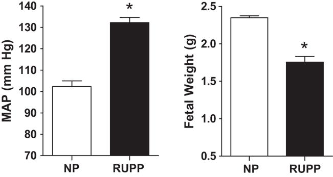
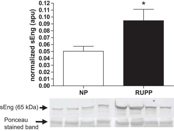
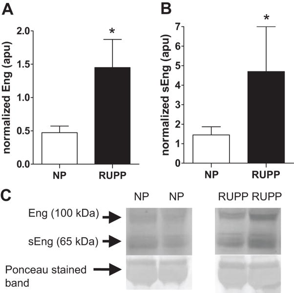
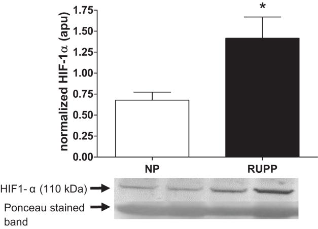
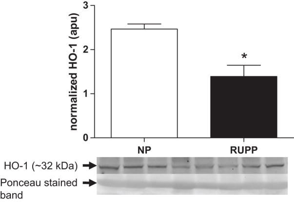
Recent clinical studies indicate that an excess of angiostatic factors, such as soluble endoglin (sEng), is related to the occurrence of preeclampsia. Although recent clinical studies report that sEng is increased in preeclamptic women, the mechanisms underlying its overexpression remain unclear. Evidence suggests that hypoxia and induction of heme oxygenase-1 have opposing effects on sEng expression, the former stimulatory and the latter inhibitory. Hence, we hypothesized that placental ischemia because of reduced uterine perfusion pressure (RUPP) in the pregnant rat would increase sEng expression and decrease heme oxygenase-1. Mean arterial pressure was obtained via arterial catheter, and serum and placental proteins were measured by Western blot. Mean arterial pressure was increased (132+/-3 mm Hg versus 102+/-2 mm Hg; P<0.001), and fetal (2.35+/-0.05 g versus 1.76+/-0.08 g; P<0.001) and placental weight were decreased (0.47+/-0.04 g versus 0.58+/-0.03 g; P<0.01) in the RUPP compared with normal pregnant controls. Serum sEng (0.10+/-0.02 arbitrary pixel units [apu] versus 0.05+/-0.01 apu; P<0.05) and placental endoglin (4.7+/-2.3 apu versus 1.45+/-0.42 apu; P<0.05) were increased along with placental hypoxia inducible factor-1 alpha (1.42+/-0.25 apu versus 0.68+/-0.09 apu; P<0.05) expression in the RUPP versus the normal pregnant dams. Placental HO-1 (1.4+/-0.3 apu versus 2.5+/-0.1 apu; P<0.05) expression decreased in the RUPP compared with normal pregnant dams. The present findings support our hypothesis that placental ischemia because of RUPP increases the expression of sEng and shifts the balance of angiogenic factors in the maternal circulation toward an angiostatic state. The present study provides further evidence that placental ischemia is a strong in vivo stimulus of angiostatic factors during pregnancy.
Conflict of interest statement
None.
Figures
References
-
- Sibai B, Dekker G, Kupferminc M. Pre-eclampsia. Lancet. 2005;365:785–799. - PubMed
-
- Roberts JM, Pearson G, Cutler J, Lindheimer M. Summary of the NHLBI Working Group on Research on Hypertension During Pregnancy. Hypertension. 2003;41:437–445. - PubMed
-
- Roberts JM, Taylor RN, Musci TJ, Rodgers GM, Hubel CA, McLaughlin MK. Preeclampsia: an endothelial cell disorder. Am J Obstet Gynecol. 1989;161:1200–1204. - PubMed
-
- Redman CW. Current topic: pre-eclampsia and the placenta. Placenta. 1991;12:301–308. - PubMed
-
- Masuyama H, Nakatsukasa H, Takamoto N, Hiramatsu Y. Correlation between Soluble Endoglin, Vascular Endothelial Growth Factor Receptor-1, and Adipocytokines in Preeclampsia. Journal of Clinical Endocrinology Metabolism. 2007;92:2672–2679. - PubMed
Publication types
MeSH terms
Substances
Grants and funding
LinkOut - more resources
Full Text Sources
Medical
Molecular Biology Databases
