. 2008 Dec 15;183(6):1061-74.
doi: 10.1083/jcb.200804003.

Assembly of the PtdIns 4-kinase Stt4 complex at the plasma membrane requires Ypp1 and Efr3

Affiliations

Assembly of the PtdIns 4-kinase Stt4 complex at the plasma membrane requires Ypp1 and Efr3

Dan Baird et al. J Cell Biol. .

Abstract

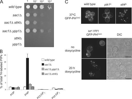
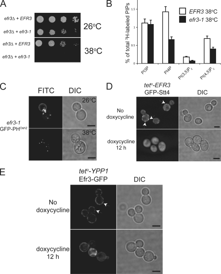
The phosphoinositide phosphatidylinositol 4-phosphate (PtdIns4P) is an essential signaling lipid that regulates secretion and polarization of the actin cytoskeleton. In Saccharomyces cerevisiae, the PtdIns 4-kinase Stt4 catalyzes the synthesis of PtdIns4P at the plasma membrane (PM). In this paper, we identify and characterize two novel regulatory components of the Stt4 kinase complex, Ypp1 and Efr3. The essential gene YPP1 encodes a conserved protein that colocalizes with Stt4 at cortical punctate structures and regulates the stability of this lipid kinase. Accordingly, Ypp1 interacts with distinct regions on Stt4 that are necessary for the assembly and recruitment of multiple copies of the kinase into phosphoinositide kinase (PIK) patches. We identify the membrane protein Efr3 as an additional component of Stt4 PIK patches. Efr3 is essential for assembly of both Ypp1 and Stt4 at PIK patches. We conclude that Ypp1 and Efr3 are required for the formation and architecture of Stt4 PIK patches and ultimately PM-based PtdIns4P signaling.


Figures

Figure 1.
Figure 1.
Ypp1 colocalizes with Stt4 at static cortical structures at the PM. (A) Cells expressing Ypp1-GFP were grown at 26°C and examined by fluorescence microscopy. Cells shown are representative of over 200 cells observed. Arrows show regions of highly concentrated Ypp1-GFP. Bar, 4 μm. (B) Cells coexpressing GFP-Stt4 and Ypp1-mCherry were grown at 26°C and examined by confocal microscopy. As previously reported (Audhya and Emr, 2002), GFP-Stt4 localizes to cortical patches on the PM (top left) and with a similar distribution as Ypp1-mCherry (top right). An overlay of the two images (bottom left) shows overlap of the fluorescent patches. Arrows indicate regions of the most conspicuous colocalization. Bar, 4 μm. (C) Schematic representation of Stt4, Ygr198w (Ypp1), and the Ypp1 human homologue TTC7a (available from GenBank/EMBL/DDBJ under accession no. NM_020458). Stt4 is a 215-kD protein with a lipid kinase unique domain (LKU) and a PtdIns 4-kinase domain (PI4K) as its only recognizable motifs. Ypp1 is a 95-kD protein encompassing multiple TPR domains and with conserved homologues in higher species. The schematics are not to scale.
Figure 2.
Figure 2.
Stt4 directly interacts with Ypp1 to form a stable complex. (A) Fluorescent image of GFP-Stt4 (left), Ypp1-GFP (right), and their corresponding kymographs (bottom). A representative section of the cell (the demarcated region in top panels) was examined for its relative movement at the cell membrane. A fluorescent time point of the demarcated region was taken every 5 s over the course of 3 min. The spatial variance of the selected fluorescent region with regard to time is represented in the bottom image. (B) FRAP of the PtdIns4P reporter GFP-2xPHOsh2 (top and Video 3, available at http://www.jcb.org/cgi/content/full/jcb.200804003/DC1) and GFP-Stt4 (bottom and Video 2). The white partitions demarcate the regions exposed to high intensity light, a FRAP region, and a control region exposed to normal excitation. The corresponding graph quantifies the fluorescent values with regard to time. (C) Cells expressing HA-Ypp1 and GFP alone or GFP-Stt4 were grown to mid-log phase, lysed, and incubated with an anti-GFP antibody. Coimmunoprecipitated proteins were detected with an anti-HA antibody. The relative abundance of coimmunoprecipitated HA-Ypp1 is shown in the top panel compared with a sample of the input from the whole cell lysate (WCL) in the bottom panel. Note that a small region of the immunoprecipitated GFP-Stt4 is cleaved at the C terminus. (D) Truncation mutants of GST-tagged Stt4 were purified from E. coli lysates, immobilized on glutathione beads, and normalized to equal amounts. Beads containing the GST-Stt4 truncations were then incubated for 1 h with lysate from E. coli overexpressing HIS6-Ypp1. Protein that bound the GST-Stt4 fragments was probed using an anti-HIS6 antibody. The relative amount of HIS6-Ypp1 that bound each respective GST-Stt4 fragment (top) and the relative amount of the Stt4 fragments that bound the glutathione beads (bottom) are shown. Notice that the primary Stt4 fragment (736–1346) is a C-terminal degradation product missing ∼200 amino acids. C-terminal degradation products are marked with an asterisk.
Figure 3.
Figure 3.
Ypp1 functions in the PtdIns4P signaling pathway to positively regulate Stt4 kinase activity. (A) The relative growth capabilities of wild type, stt4-4, and ypp1-7 at 26 (top left) and 37°C (bottom left). The relative growth capability of ypp1-7 with overexpression of YPP1 from a 2μ plasmid, plasmid alone (pRS426), or overexpressed STT4 at 37°C (top right). Similar growth profile of stt4-4 at 37°C in the presence of vector alone or in the presence of overexpressed YPP1 (bottom right). (B) Whole cell lysate from teto-YPP1-HA cells grown in the presence or absence of doxycycline were analyzed by Western blot with an anti-HA antibody. The relative abundance of Ypp1-HA under the indicated conditions (top) and the relative abundance of a control protein, G6PDH, from the same lysate (bottom) are shown. (C) Phosphoinositide levels in teto-YPP1 cells with and without doxycycline and control R1158 in the presence of doxycycline. Data are presented as means and SD (error bars) of three independent experiments.
Figure 4.
Figure 4.
Deletion of SAC1 bypasses ypp1Δ or stt4Δ. (A) Relative growth profiles of wild type, sac1Δ, sac1Δ stt4Δ, sac1Δ ypp1Δ, stt4Δ, and ypp1Δ at 26°C. (B) Phosphoinositide levels of wild type, sac1Δ, sac1Δ stt4Δ, and sac1Δ ypp1Δ. Data are presented as means and SD (error bars) of three independent experiments. (C) Cellular localization of the PtdIns4P reporter GFP-2xPHOsh2 in wild type, pik1ts, and stt4ts (top) at the restrictive temperature. (bottom) The fluorescent localization of GFP-2xPHOsh2 in teto-YPP1 grown in the presence (bottom) or absence (top) of doxycycline for 20 h. Bars, 4 μm.
Figure 5.
Figure 5.
Stt4 and Ypp1 are codependent for proper localization and organization into PIK patches at the PM. (A) Relative abundance of GFP-Stt4 after doxycycline treatment in teto-YPP1 cells over the time course of 16 h. The relative abundance of G6PDH is shown as a control. (B) The localization profile of teto-YPP1 GFP-Stt4 in the absence (top) or presence (second panel) of doxycycline and the corresponding DIC image. The localization teto-STT4 GFP-Ypp1 in the absence (third panel) or presence (bottom) of doxycycline and the corresponding DIC images. Bars, 4 μm. (C) Localization of PM anchored Psr11-28-GFP-Ypp1 expressed under wild-type conditions (left) or in the sac1Δ stt4Δ background (right). The graph is the relative fluorescence intensity along the PM within the indicated spatial region. Arrows indicating the start and end of the fluorescent images correspond to the x axis of the graph.
Figure 6.
Figure 6.
Stt4 stabilizes Ypp1 molecules on a membrane-bound fraction of the cell. (A) Subcellular fractionation of cells expressing HA-Ypp1 (left) in a wild-type background and HA-Ypp1 distribution in the absence of SAC1 and STT4 (right). Protein component fractions in the pellet (P100; rcf of 100,000) or soluble (S100; soluble portion) fractions. Fractionation of G6PDH (soluble) and Pep13 (membrane-tethered Golgi component) are shown as controls. (B) FPLC elution profile of HA-Ypp1 isolated from the S100 portion of sac1Δ stt4Δ cells (top). Fractions were collected at the indicated elution volumes, separated by SDS-PAGE, and detected using an anti-HA antibody. The molecular mass corresponding to each elution is indicated below the blot. (C) Coimmunoprecipitation of HA-Ypp1 from yeast lysate that coexpressed GFP alone, GFP-Ypp1, or GFP-Stt4. (D) Coimmunoprecipitation of HA-Ypp1 with GFP-Ypp1 in the wild type, sac1Δ, and sac1Δ stt4Δ background. (E) FPLC sizing of E. coli–expressed and purified HIS6-Ypp1 using a Superdex 200 analytical column.
Figure 7.
Figure 7.
Efr3 is a component of PIK patches at the PM. (A) Tetrad dissection of diploid yeast with a deleted chromosomal copy of efr3. (B) Full-length and truncation mutants of GST-tagged Efr3 were purified from E. coli lysates, immobilized on glutathione beads, and normalized to equal amounts. Beads containing the GST-Efr3 constructs were then incubated for 1 h with yeast lysate expressing Ypp1-GFP. Protein that bound the GST-Efr3 fragments was probed using an anti-GFP antibody. (C) Subcellular fractionation of Efr3-GFP from wild-type cell extracts. (D) Cells coexpressing Efr3-GFP and Ypp1-mCherry were grown at 26°C and examined by fluorescence microscopy. Efr3-GFP localizes to cortical patches on the PM (left) and with a similar distribution as Ypp1-mCherry (right). Arrows on the Efr3-GFP image indicate regions of colocalization. Bar, 4 μm.
Figure 8.
Figure 8.
Efr3 recruits PIK patch components to the PM. (A) Growth assay of EFR3 wild type and efr3-1 temperature-sensitive yeast at 26 and 38°C. (B) Phosphoinositide levels of EFR3 and efr3-1 at 38°C. Data are presented as means and SD (error bars) of two independent experiments. (C) Cellular localization of the PtdIns4P reporter GFP-2xPHOsh2 in efr3-1 yeast at the permissive (top) and restrictive (bottom) temperatures. Bars, 4 μm. (D) The localization profile of teto-EFR3 GFP-Stt4 in the absence (top) or presence (bottom) of doxycycline and the corresponding DIC image. Arrows indicate PIK patch localization. Bars, 4 μm. (E) The localization profile of teto-YPP1 Efr3-GFP in the absence (top) or presence (bottom) of doxycycline and the corresponding DIC images. Arrows indicate PIK patch localization. Bars, 4 μm.
Figure 9.
Figure 9.
Organization of Stt4 PIK patches at the PM. Ypp1 is a multivalent linker of Stt4 molecules ensuring the stability of the lipid kinase and the organization of the PIK patch. Efr3 anchors the Stt4–Ypp1 complex at the PM by binding Ypp1. TMD, transmembrane domain; LKUD, lipid kinase unique domain; PI4K, PtdIns 4-kinase domain.

Similar articles

Cited by

References

    1. Aronov, S., and J.E. Gerst. 2004. Involvement of the late secretory pathway in actin regulation and mRNA transport in yeast. J. Biol. Chem. 279:36962–36971. - PubMed
    1. Audhya, A., and S.D. Emr. 2002. Stt4 PI 4-kinase localizes to the plasma membrane and functions in the Pkc1-mediated MAP kinase cascade. Dev. Cell. 2:593–605. - PubMed
    1. Audhya, A., M. Foti, and S.D. Emr. 2000. Distinct roles for the yeast phosphatidylinositol 4-kinases, Stt4p and Pik1p, in secretion, cell growth, and organelle membrane dynamics. Mol. Biol. Cell. 11:2673–2689. - PMC - PubMed
    1. Blatch, G.L., and M. Lassle. 1999. The tetratricopeptide repeat: a structural motif mediating protein-protein interactions. Bioessays. 21:932–939. - PubMed
    1. Brickner, J.H., and P. Walter. 2004. Gene recruitment of the activated INO1 locus to the nuclear membrane. PLoS Biol. 2:e342. - PMC - PubMed

Publication types

MeSH terms

Substances