. 2009 Apr 30;113(18):4381-90.
doi: 10.1182/blood-2008-08-174748. Epub 2008 Dec 15.

Structure of the Notch1-negative regulatory region: implications for normal activation and pathogenic signaling in T-ALL

Affiliations

Structure of the Notch1-negative regulatory region: implications for normal activation and pathogenic signaling in T-ALL

Wendy R Gordon et al. Blood. .

Abstract

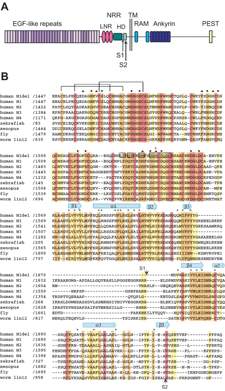
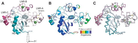
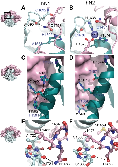
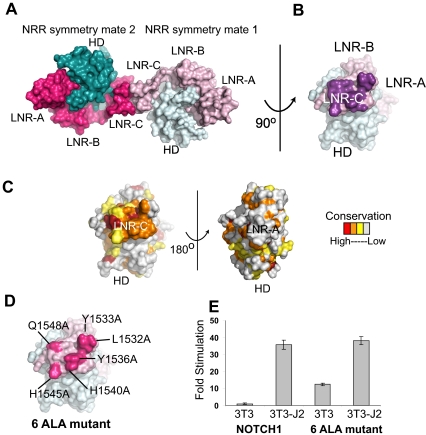
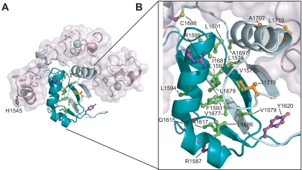
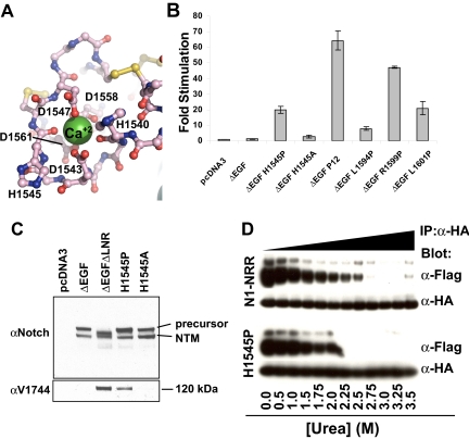
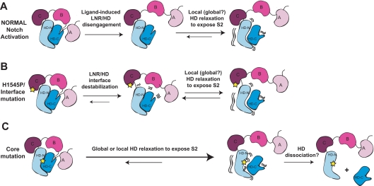
Proteolytic resistance of Notch prior to ligand binding depends on the structural integrity of a negative regulatory region (NRR) of the receptor that immediately precedes the transmembrane segment. The NRR includes the 3 Lin12/Notch repeats and the juxtamembrane heterodimerization domain, the region of Notch1 most frequently mutated in T-cell acute lymphoblastic leukemia lymphoma (T-ALL). Here, we report the x-ray structure of the Notch1 NRR in its autoinhibited conformation. A key feature of the Notch1 structure that maintains its closed conformation is a conserved hydrophobic plug that sterically occludes the metalloprotease cleavage site. Crystal packing interactions involving a highly conserved, exposed face on the third Lin12/Notch repeat suggest that this site may normally be engaged in intermolecular or intramolecular protein-protein interactions. The majority of known T-ALL-associated point mutations map to residues in the hydrophobic interior of the Notch1 NRR. A novel mutation (H1545P), which alters a residue at the crystal-packing interface, leads to ligand-independent increases in signaling in reporter gene assays despite only mild destabilization of the NRR, suggesting that it releases the autoinhibitory clamp on the heterodimerization domain imposed by the Lin12/Notch repeats. The Notch1 NRR structure should facilitate a search for antibodies or compounds that stabilize the autoinhibited conformation.


Figures

Figure 1
Figure 1
Domain organization and multiple sequence alignment. (A) Domain organization of human Notch1. The NRR consists of the LNR and HD domains. Adapted from Gordon et al. (B) Sequence alignment of the NRR region of various Notch receptors, colored according to sequence conservation: red indicates absolutely conserved; orange, highly conserved (defined by ClustalW strong conservation groups and/or > 80% sequence identity); yellow, moderately conserved (defined by ClustalW weak conservation groups or > 50% sequence identity); and white, nonconserved. Amino acid residues of special importance are denoted as follows: side-chain and main-chain Ca++-coordinating residues, circles and triangles, respectively; residues mutated in Notch1 in T-ALL, asterisks. Residues from LNR-C engaged in crystal lattice contacts are boxed. Representative disulfide connectivity is shown for LNR-A and HD secondary structural elements are represented by arrows (beta strands) and cylinders (alpha helices).
Figure 2
Figure 2
Overall structure of the human Notch1 NRR and comparison with the human Notch2 NRR. (A) Overall structure of the human Notch1 NRR, shown in ribbon representation. Shades of pink and purple represent LNR modules; shades of light and dark cyan, HD domain (on N- and C-terminal sides of the furin cleavage loop, respectively). Disulfide bonds are rendered as yellow sticks, and calcium ions as green spheres. Arrows denote positions of the S1 and S2 cleavage sites. (B) Ribbon representation of the Notch1 NRR colored according to the root mean square deviation (RMSD, in angstroms) between corresponding alpha carbon atoms of the Notch1 and Notch2 structures. Colors are assigned on a sliding scale from blue (RMSD = 0) to red (RMSD = 7.25 Å). Residues present in Notch1 that are absent in Notch2 were set to the maximum RMSD of the range. (C) Overlay of the backbone traces of the Notch1 (colored as in A) and Notch2 (gray) NRRs.
Figure 3
Figure 3
Structural divergence between Notch1 and Notch2 NRRs. (A,B) Convergence of HD-N, HD-C, and LNR-C is mediated by a hydrogen bond in the Notch1 NRR (A) and a Zn2+ coordination site in the Notch2 NRR (B). (C,D) Comparison of interactions stabilizing helix 1 in the Notch1 and Notch2 NRRs. In Notch1, there is an intrahelical salt bridge and the helix is anchored to LNR-C via a single salt bridge. In Notch2, there are several electrostatic interactions between the helix and LNR-C (D). (E,F) The LNR A-B linker of the Notch1 (E) and Notch2 (F) NRRs masks the metalloprotease cleavage site. In each structure, a 3-residue sequence from the linker occludes the S2 site, even though 2 of the 3 amino acid residues comprising the protective plug are not conserved (see “Interdomain interactions” for details). Black labels identify residues participating in the interactions discussed, whereas blue labels identify residues that form analogous interactions in the other receptor.
Figure 4
Figure 4
Conserved residues at a Notch1 crystal packing interface. (A) The Notch1 NRR and a symmetry mate are depicted in molecular surface representation. The LNR domains are colored in light and dark pink and the HD domains colored in light and dark cyan in molecules 1 and 2, respectively. (B) Molecule one surface after a counterclockwise 90-degree rotation about the axis shown. In this view, the surface-exposed face of LNR-C is facing outward. Amino acid residues involved in the crystal contact interface are colored purple. (C) Surface of the Notch1 NRR colored according to amino acid conservation using the scale shown (as described in the legend for Figure 1B). (D) Surface of the Notch1 NRR illustrating the 6 crystal contact residues that have been mutated to Ala (hot pink). (E) Effect of mutating the LNR-C crystal contact interface in Notch reporter gene assays. Signaling in coculture assays with NIH 3T3 cells alone or stably expressing the ligand Jagged-2 was examined using chimeric full-length receptors in which the RAM and ANK domains of NOTCH1 were replaced with the DNA-binding domain of the transcription factor GAL4 (“Luciferase reporter/urea sensitivity assays”). Firefly luciferase activity was normalized to the internal Renilla control and expressed relative to the reporter gene activity of the unmutated chimeric receptor cocultured with NIH 3T3 cells alone. Error bars represent the SE of the 3 replicate measurements made for each experimental condition.
Figure 5
Figure 5
Mapping of T-ALL tumor-associated mutations onto the Notch1 NRR. (A) Structural representation highlighting T-ALL mutations. Side chains of residues mutated in T-ALL patients are shown in ball-and-stick form. Residues are colored according to mutation site: core (green), interface (orange), or partially exposed (purple). (B) Close-up view of mutations in the HD domain.
Figure 6
Figure 6
Characterization of the H1545P mutation in LNR-C. (A) Structure of the region surrounding H1545. Calcium coordinating residues are shown in ball-and-stick representation and are colored by atom type (gray indicates carbon; red: oxygen; and blue: nitrogen). Disulfide bonds are yellow, and the calcium ion is a green sphere. (B) Reporter gene assay for ligand-independent Notch1 activation. H1545P and several other well-characterized T-ALL–associated mutations were tested for their ability to induce reporter gene transcription in the context of a Notch1 polypeptide lacking the EGF-like ligand binding repeats (ΔEGF). The ΔEGF forms of the receptors were transiently transfected into U2OS cells together with a plasmid encoding a luciferase reporter gene under control of 4 iterated CSL binding sites, and an internal control plasmid expressing Renilla luciferase. Firefly luciferase activities were normalized to the internal Renilla control and expressed relative to the activity produced by the unmutated ΔEGF form of the receptor, which was assigned a relative value of 1. Error bars represent the SE of the 3 replicate measurements made for each experimental condition. (C) The H1545P mutation confers ligand-independent proteolytic sensitivity. U2OS cells were transfected with plasmids encoding the indicated Notch1 receptor variants. Western blots with antibodies specific for the intracellular portion of human Notch1 (top panel) and for the S3-cleaved product (bottom panel) are shown. (D) Effect of the H1545P mutation on sensitivity to urea-induced subunit dissociation. Conditioned media from HEK 293T cells expressing epitope-tagged Notch1 NRR minireceptors were immunoprecipitated with α-HA coupled beads followed by incubation in buffer containing different concentrations of urea for 30 minutes (0 to 3.5 M). Subunit dissociation was evaluated by SDS–polyacrylamide gel electrophoresis (PAGE) followed by Western blot analysis. The N-terminal and C-terminal subunits were detected with α-FLAG and α-HA antibodies, respectively.
Figure 7
Figure 7
Models for normal Notch activation and aberrant activation by T-ALL mutations. (A) Ligand-mediated Notch activation. Ligand binding triggers a conformational movement that first disengages the LNR and HD domains. Without the stabilizing interactions provided by the LNR domain, the HD domain then relaxes locally or globally to expose the S2 site. (B) H1545P and other interface mutations promote disengagement of the LNR/HD interface. This step then allows local or global HD relaxation to expose the S2 site in a manner analogous to ligand binding. (C) Core mutations directly destabilize the HD domain, precluding stable interaction with the LNR domain and promoting exposure of S2. Extremely destabilizing mutations such as those of class 1A may lead to complete HD dissociation.

References

    1. Bray SJ. Notch signalling: a simple pathway becomes complex. Nat Rev Mol Cell Biol. 2006;7:678–689. - PubMed
    1. Blaumueller CM, Qi H, Zagouras P, Artavanis-Tsakonas S. Intracellular cleavage of Notch leads to a heterodimeric receptor on the plasma membrane. Cell. 1997;90:281–291. - PubMed
    1. Logeat F, Bessia C, Brou C, et al. The Notch1 receptor is cleaved constitutively by a furin-like convertase. Proc Natl Acad Sci U S A. 1998;95:8108–8112. - PMC - PubMed
    1. Kopan R, Schroeter EH, Weintraub H, Nye JS. Signal transduction by activated mNotch: importance of proteolytic processing and its regulation by the extracellular domain. Proc Natl Acad Sci U S A. 1996;93:1683–1688. - PMC - PubMed
    1. Sanchez-Irizarry C, Carpenter AC, Weng AP, Pear WS, Aster JC, Blacklow SC. Notch subunit heterodimerization and prevention of ligand-independent proteolytic activation depend, respectively, on a novel domain and the LNR repeats. Mol Cell Biol. 2004;24:9265–9273. - PMC - PubMed

Publication types

MeSH terms