. 2008 Dec 23;105(51):20333-8.
doi: 10.1073/pnas.0809045105. Epub 2008 Dec 15.

A postnatal switch of CELF and MBNL proteins reprograms alternative splicing in the developing heart

Affiliations

A postnatal switch of CELF and MBNL proteins reprograms alternative splicing in the developing heart

Auinash Kalsotra et al. Proc Natl Acad Sci U S A. .

Abstract

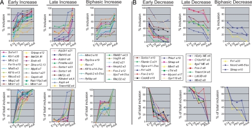
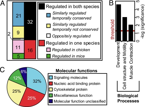
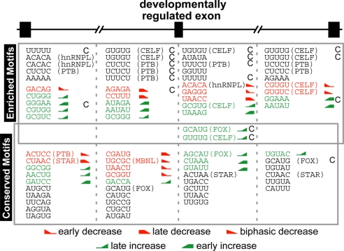
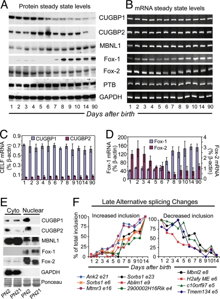
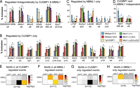
From a large-scale screen using splicing microarrays and RT-PCR, we identified 63 alternative splicing (AS) events that are coordinated in 3 distinct temporal patterns during mouse heart development. More than half of these splicing transitions are evolutionarily conserved between mouse and chicken. Computational analysis of the introns flanking these splicing events identified enriched and conserved motifs including binding sites for CUGBP and ETR-3-like factors (CELF), muscleblind-like (MBNL) and Fox proteins. We show that CELF proteins are down-regulated >10-fold during heart development, and MBNL1 protein is concomitantly up-regulated nearly 4-fold. Using transgenic and knockout mice, we show that reproducing the embryonic expression patterns for CUGBP1 and MBNL1 in adult heart induces the embryonic splicing patterns for more than half of the developmentally regulated AS transitions. These findings indicate that CELF and MBNL proteins are determinative for a large subset of splicing transitions that occur during postnatal heart development.


Conflict of interest statement

The authors declare no conflict of interest.

Figures

Fig. 1.
Fig. 1.
Subsets of AS transitions are coregulated during specific times of mouse heart development. Total RNA was isolated from 6–20 pooled hearts at the indicated time points. RT-PCR analysis was carried out for 63 AS events. Data are expressed as the percentage inclusion (Upper) and as the percentage of total change (Lower) for variable regions that show increased (A) or decreased (B) inclusion during development. Alternative exons are numbered according to Ensembl, and sequences of variable regions are presented in Table S2. In at least 2 independent assays for all E14 and adult samples, the standard deviation was <5 percentage points.
Fig. 2.
Fig. 2.
Most of the splicing transitions regulated during mouse heart development undergo similar transitions during chicken heart development. (A) Conservation of AS in mouse and chicken heart development. (B) Gene ontology of 30 events conserved between chicken and mouse. Enriched biological processes are plotted on a −log (P value) scale with the threshold set to 1.3 [log (0.05)]. (C) Molecular functions assigned to the genes falling in the most significantly enriched process “Development” (P < 0.000005).
Fig. 3.
Fig. 3.
Motifs significantly enriched (Upper) and/or conserved (P < 0.001) among 8 mammalian species (Lower) in the 4 flanking 250-nt intronic regions of developmentally regulated mouse exons. Motifs that are significantly conserved and/or enriched that also exhibit a significant (P ≤ 0.05) association with specific temporal transitions in the regression analysis are indicated in red (decreasing inclusion) or green (increasing inclusion) with icons indicating the transition pattern. Motifs in black are significantly conserved or enriched but not significant the regression analysis. “C” indicates that the motif is also significantly enriched among the developmentally regulated chicken exons. The 5 most significant pentamers are shown (see Table S5, Table S6, Table S7, and Table S8 for the complete lists of motifs). Motifs resembling known binding sites of splicing factors (Table S10) are annotated. Fox (GCAUG) and CELF (GUGUG) motifs were significant in all analyses being enriched in mouse and chicken, conserved in mammals, and associated with a specific temporal pattern.
Fig. 4.
Fig. 4.
Postnatal expression of CELF, Fox, and MBNL proteins correlate with AS transitions. (A) Steady-state protein levels. (B) Steady-state mRNA levels by RT-PCR. (C and D) Steady-state mRNA levels determined by real time RT-PCR (TaqMan). (E) Nuclear and cytoplasmic distributions of CUGBP1, CUGBP2, Fox-1, Fox-2, and MBNL1. The distribution of Fox-1 and 2 or GAPDH demonstrate the clean separation of nuclear and cytoplasmic fractions. (F) A tight postnatal time course demonstrates a correlation between the timing of postnatal AS transitions and changes in CELF, Fox, and MBNL expression.
Fig. 5.
Fig. 5.
A subset of postnatal splicing transitions are regulated by CUGBP1 and/or MBNL1. Forty-four AS events were tested in the hearts of TgCUGBP1 and Mbnl1ΔE3/ΔE3 mice and their respective littermate controls. (A–D) Splicing events were found to be regulated antagonistically by CUGBP1 and MBNL1 (A), regulated by CUGBP1 only (B), regulated by MBNL1 only (C), or CUGBP1 and MBNL1 independent (D). Each bar shows the mean ± SD for the percent inclusion of the specified variable region. Statistical analysis was done by using 1-way ANOVA, followed by Tukey's multiple-range test (P < 0.05). *, significantly different from E14. †, significantly different from wild-type littermates. (E–H) Computational analysis for enrichment of CELF- and MBNL-binding motifs in flanking introns of CUGBP1- and MBNL1-regulated exons. To the right of each plot are heat maps representing the P values for significant enrichment of indicated motifs in positions 12–250 of the upstream intron (upIn1), positions −250 to −31 of the upstream intron (upIn), positions 12 to 250 of the downstream intron (dnIn), and positions −250 to −31 of the downstream intron (dnIn1).

References

    1. Graveley BR. Alternative splicing: Increasing diversity in the proteomic world. Trends Genet. 2001;17:100–107. - PubMed
    1. Johnson JM, et al. Genome-wide survey of human alternative pre-mRNA splicing with exon junction microarrays. Science. 2003;302:2141–2144. - PubMed
    1. Wang GS, Cooper TA. Splicing in disease: Disruption of the splicing code and the decoding machinery. Nat Rev Genet. 2007;8:749–761. - PubMed
    1. Gabut M, Chaudhry S, Blencowe BJ. SnapShot: The splicing regulatory machinery. Cell. 2008;133:192–e1. - PubMed
    1. Matlin AJ, Clark F, Smith CW. Understanding alternative splicing: Towards a cellular code. Nat Rev Mol Cell Biol. 2005;6:386–398. - PubMed

Publication types