. 2008 Dec 23;105(51):20505-10.
doi: 10.1073/pnas.0811201106. Epub 2008 Dec 15.

Etifoxine improves peripheral nerve regeneration and functional recovery

Affiliations

Etifoxine improves peripheral nerve regeneration and functional recovery

Christelle Girard et al. Proc Natl Acad Sci U S A. .

Abstract

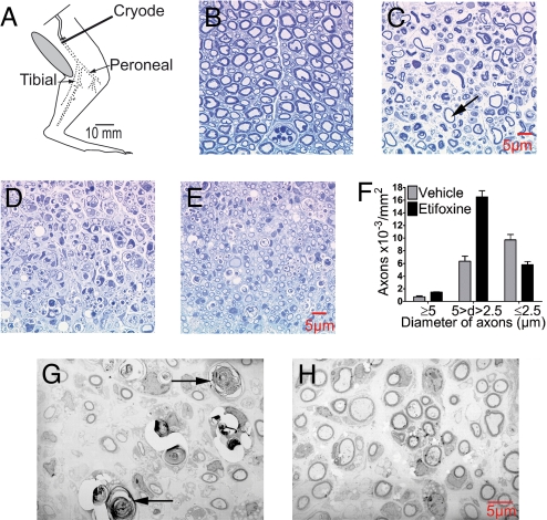
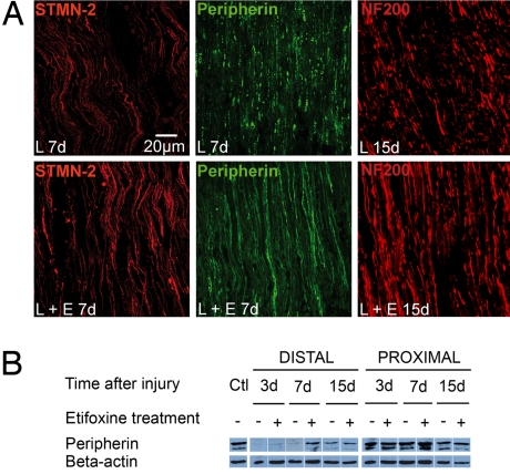
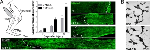
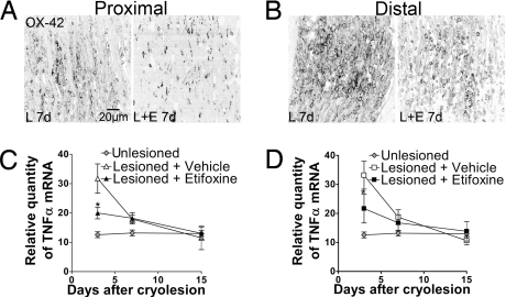
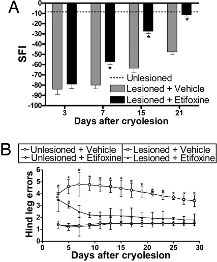
Peripheral nerves show spontaneous regenerative responses, but recovery after injury or peripheral neuropathies (toxic, diabetic, or chronic inflammatory demyelinating polyneuropathy syndromes) is slow and often incomplete, and at present no efficient treatment is available. Using well-defined peripheral nerve lesion paradigms, we assessed the therapeutic usefulness of etifoxine, recently identified as a ligand of the translocator protein (18 kDa) (TSPO), to promote axonal regeneration, modulate inflammatory responses, and improve functional recovery. We found by histologic analysis that etifoxine therapy promoted the regeneration of axons in and downstream of the lesion after freeze injury and increased axonal growth into a silicone guide tube by a factor of 2 after nerve transection. Etifoxine also stimulated neurite outgrowth in PC12 cells, and the effect was even stronger than for specific TSPO ligands. Etifoxine treatment caused a marked reduction in the number of macrophages after cryolesion within the nerve stumps, which was rapid in the proximal and delayed in the distal nerve stumps. Functional tests revealed accelerated and improved recovery of locomotion, motor coordination, and sensory functions in response to etifoxine. This work demonstrates that etifoxine, a clinically approved drug already used for the treatment of anxiety disorders, is remarkably efficient in promoting acceleration of peripheral nerve regeneration and functional recovery. Its possible mechanism of action is discussed, with reference to the neurosteroid concept. This molecule, which easily enters nerve tissues and regulates multiple functions in a concerted manner, offers promise for the treatment of peripheral nerve injuries and axonal neuropathies.


Conflict of interest statement

The authors declare no conflict of interest.

Figures

Fig. 1.
Fig. 1.
Cryolesion of the rat sciatic nerve resulted in the complete degeneration of nerve fibers. Treatment with etifoxine promoted nerve fiber regeneration at the lesion site. (A) Diagram illustrating cryolesion of the rat sciatic nerve. Dotted line indicates nerve undergoing Wallerian degeneration distal to the freeze lesion. Semithin sections (B–E) and ultrathin sections (G and H). (B) Intact rat sciatic nerve. (C) Complete destruction of myelinated axons 3 days after cryolesion. Arrow indicates remaining myelin sheath surrounding an empty space left by a degenerated axon. No spared fibers were observed. (D) Regenerated nerve fibers 15 days after cryolesion in rats treated with vehicle and (E) in rats treated with etifoxine. (F) Fifteen days after cryolesion, the number of regenerated medium-sized axons (diameter 2.5–5 μm) surrounded by myelin sheaths was increased by etifoxine. (G) Regenerated nerve fibers by electron microscopy in rats treated with vehicle (arrows indicate degenerated axons) and (H) in rats treated with etifoxine at 15 days.
Fig. 2.
Fig. 2.
Etifoxine treatment accelerated the regeneration of axons expressing either STMN-2, peripherin, or NF200 at the site of freeze injury within the distal stump. Immunoreactive fibers were observed by confocal microscopy. (A) STMN-2–immunoreactive axons (red) and peripherin-immunoreactive axons (green) 7 days after cryolesion (7d) and NF200-immunoreactive fibers 15 days after cryolesion (15d). Lesioned sciatic nerves were treated with vehicle (L) or etifoxine (L + E). (B) Western blot analysis of peripherin, normalized to β-actin. Ctl = unlesioned control.
Fig. 3.
Fig. 3.
Etifoxine treatment promoted axonal extension after sciatic nerve transection and neurite outgrowth in PC12 cells. (A) Diagram illustrating transection of the rat sciatic nerve and installation of a silicone tube (dotted line indicates transected nerve undergoing Wallerian degeneration). STMN-2–immunoreactive axons (green) showed the increase of the elongation rate from the proximal stump into the tube, by more than 2-fold, 7 days (7d) and 10 days (10d) after surgery. The dotted white line delimits the extension of the regenerated axons. The maximal extension of nerve fibers into the tube was quantified at 7, 10, and 15 days after sciatic nerve transection. (B) Etifoxine (E) (20 μM) enhanced neurite extension in PC12 cells grown for 72 h in the presence of 10 ng/ml NGF.
Fig. 4.
Fig. 4.
Influence of etifoxine (E) treatment on (A and B) the number of activated macrophages and (C and D) mRNA expression of the cytokine TNF-α in the regenerating sciatic nerve. The density of OX-42–immunoreactive macrophages is shown at 7 days (7d) after cryolesion (L) in the nerve endings (A) proximal and (B) distal to the lesion site. Relative mRNA levels were analyzed by real-time RT-PCR within the nerve stumps (C) proximal and (D) distal to the injury site (n = 5). *, P < 0.01 vs. vehicle-treated lesioned nerves by Tukey's tests after two-way ANOVA (treatment × time after cryolesion).
Fig. 5.
Fig. 5.
Etifoxine treatment accelerated and improved functional recovery after cryolesion of the sciatic nerve. (A) Locomotion was assessed by the walking track test, and footprints were monitored at 3, 7, 15, and 21 days after cryolesion and in unlesioned control rats. Improved recovery of locomotion in etifoxine-treated animals was followed by SFI indexes, calculated according to the Dijkstra method (39). (B) Recovery of fine motor coordination was assessed in the Locotronic device by recording the number of hind footfalls. For the results of both walking track test and Locotronic device, two-way ANOVA with treatment and time after cryolesion as factors revealed both factors and their interaction to be significant at least at the 0.01 level. *, P < 0.01 vs. unlesioned rats by Tukey's tests after two-way ANOVA (n = 35; mean ± SEM).

References

    1. Barakat-Walter I. Role of thyroid hormones and their receptors in peripheral nerve regeneration. J Neurobiol. 1999;40:541–559. - PubMed
    1. Schumacher M, et al. Progesterone synthesis and myelin formation in peripheral nerves. Brain Res Rev. 2001;37:343–359. - PubMed
    1. Gold BG, Udina E, Bourdette D, Navarro X. Neuroregenerative and neuroprotective actions of neuroimmunophilin compounds in traumatic and inflammatory neuropathies. Neurol Res. 2004;26:371–380. - PubMed
    1. Melcangi RC, Garcia-Segura LM. Therapeutic approaches to peripheral neuropathy based on neuroactive steroids. Expert Rev Neurother. 2006;6:1121–1125. - PubMed
    1. Papadopoulos V, et al. Translocator protein (18kDa): New nomenclature for the peripheral-type benzodiazepine receptor based on its structure and molecular function. Trends Pharmacol Sci. 2006;27:402–409. - PubMed

Publication types

LinkOut - more resources