. 2009 Jan;119(1):61-9.
doi: 10.1172/JCI35997. Epub 2008 Dec 15.

IL-17A and IL-17F do not contribute vitally to autoimmune neuro-inflammation in mice

Affiliations

IL-17A and IL-17F do not contribute vitally to autoimmune neuro-inflammation in mice

Stefan Haak et al. J Clin Invest. 2009 Jan.

Abstract

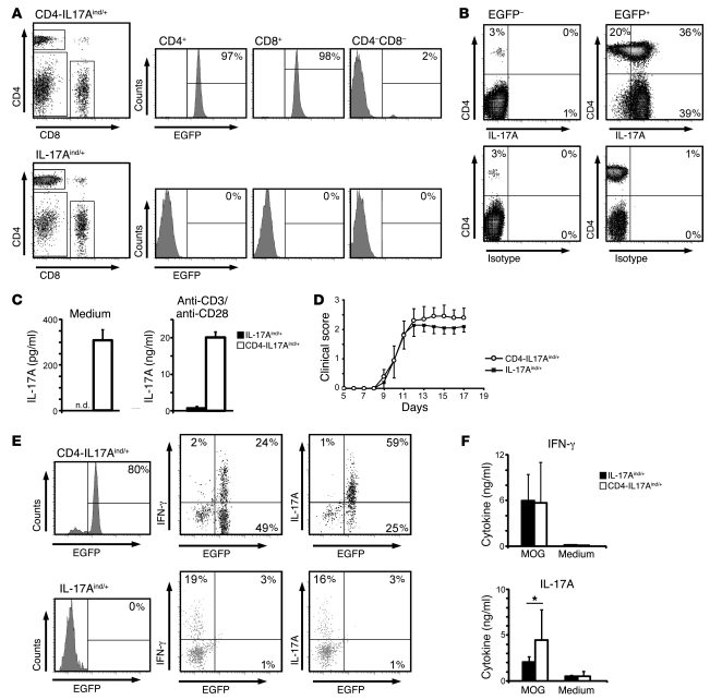
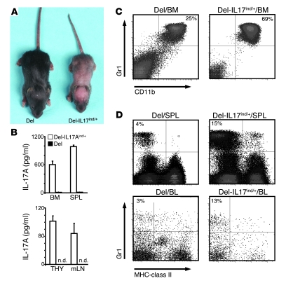
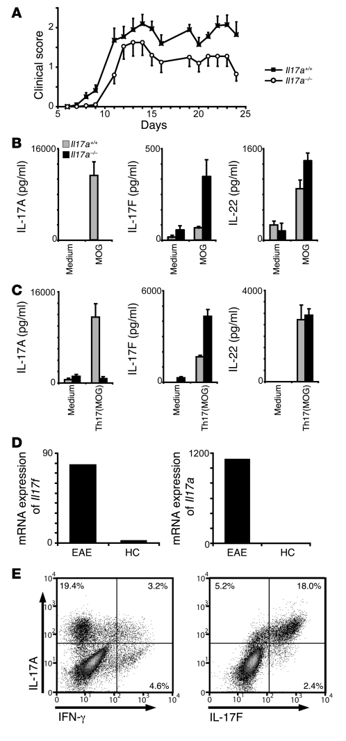
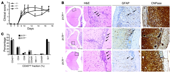
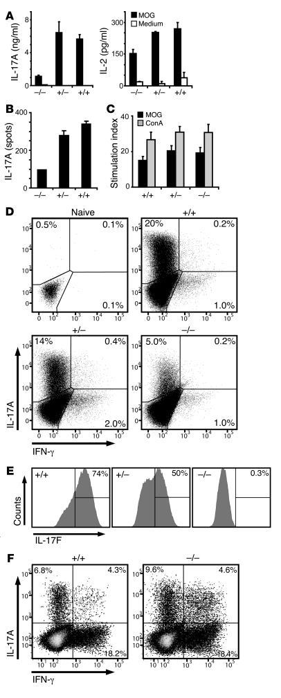
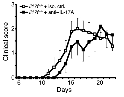
The clear association of Th17 cells with autoimmune pathogenicity implicates Th17 cytokines as critical mediators of chronic autoimmune diseases such as EAE. To study the impact of IL-17A on CNS inflammation, we generated transgenic mice in which high levels of expression of IL-17A could be initiated after Cre-mediated recombination. Although ubiquitous overexpression of IL-17A led to skin inflammation and granulocytosis, T cell-specific IL-17A overexpression did not have a perceptible impact on the development and health of the mice. In the context of EAE, neither the T cell-driven overexpression of IL-17A nor its complete loss had a major impact on the development of clinical disease. Since IL-17F may be able to compensate for the loss of IL-17A, we also generated IL-17F-deficient mice. This strain was fully susceptible to EAE and displayed unaltered emergence and expansion of autoreactive T cells during disease. To eliminate potential compensatory effects of either cytokine, we treated IL-17F-deficient mice with antagonistic monoclonal antibodies specific for IL-17A and found again only a minimal beneficial impact on disease development. We conclude therefore that both IL-17A and IL-17F, while prominently expressed by an encephalitogenic T cell population, may only marginally contribute to the development of autoimmune CNS disease.


Figures

Figure 1
Figure 1. IL-17A overexpression does not exacerbate EAE.
(A) LNs from CD4-IL17Aind/+ and IL-17Aind/+ littermates were isolated and stained for CD4 and CD8 coreceptors. EGFP expression is depicted in the histograms after indicated gating. Percentages of gated cells are shown. (B) Splenocytes from naive CD4-IL17Aind/+ mice were restimulated in the presence of Brefeldin A and subsequently stained for CD4 and IL-17A. Percentages of cells in the quadrants are indicated in the corners after gating was performed on all EGFP (plots on the left) or all EGFP+ (plots on the right) cells. Data shown are representative of 3 independent experiments. (C) FACS-sorted CD4+ T cells (2 × 10⁵) were cultured for 24 hours in the presence or absence of anti-CD3 and anti-CD28, after which IL-17A secretion was measured by flow cytomix assay. Error bars represent mean ± SD. n.d., not detectable. (D) Clinical scores after MOG35–55-induced EAE are not significantly altered by increased IL-17A expression in CD4-IL17Aind/+ and IL-17Aind/+ littermates. Error bars represent mean ± SEM. Data shown represent 1 out of 3 independent experiments. (E) Lymphocytes isolated from the diseased EAE brain and spinal cord at day 14 from CD4-IL17Aind/+ and IL-17Aind/+ littermates were restimulated and surface stained for CD4 and examined for EGFP expression. Further staining for IL-17A and IFN-γ was performed. Percentages of EGFP+IL-17A+ or EGFP+IFN-γ+ are given in the quadrant corners. Plots shown are gated on CD4+ CNS-derived T cells. (F) At peak disease, mononuclear infiltrates were isolated from inflamed CNS extracts from either CD4-IL17Aind/+ or IL-17Aind/+ mice. Cellular extracts were cultured for 2 days in the presence of 20 μg/ml MOG peptide, after which cytokine secretions were measured by flow cytomix. Error bars represent mean ± SD and significance is shown where relevant. *P = 0.039, Student’s t test.
Figure 2
Figure 2. Systemic overexpression of IL-17A in Del-IL17Aind/+ leads to granulocytosis and anemia.
(A) Crossing the IL-17Aind allele to the delete-cre strain yields mice called Del-IL17Aind/+. Skin inflammation, stunted growth, and failure to thrive is a consistent phenotype in all mice observed when compared with delete-cre littermate controls (Del). (B) Single-cell suspensions from bone marrow, spleen (SPL), thymus (THY), and mesenteric LNs (mLN) were placed in unstimulated culture for 24 hours, after which IL-17A secretion was assayed by ELISA. Error bars represent mean ± SEM. (C) Bone marrow cells were isolated from Del-IL17Aind/+ mice and littermate controls and surface stained for Gr1 and CD11b. Percentages of gated granulocytes are shown in quadrant corners. (D) Isolated spleen cells and PBMCs from the indicated genotypes were stained for MHC-class II and Gr1. Percentages of gated cells are shown. (AD) Data shown are representative of at least 2 independent experiments.
Figure 3
Figure 3. IL-17A is redundant in the induction of EAE, which could be due to a compensatory increase of IL-17F production.
(A) EAE was induced in Il17a–/– and Il17a+/+ mice by immunization with MOG35–55/CFA. The graph shows the development of EAE according to clinical scores (n = 10; SEM as indicated) in 1 out of 2 independent experiments. (B and C) Th17-cytokine profile measured by ELISA of splenocytes isolated from mice with active EAE and restimulated with MOG35–55 with (C) or without (B) the addition of Th17 polarizing conditions for 2 days. Error bars represent mean ± SEM. (D) Comparative mRNA expression analysis of Il17f and Il17a in the cerebellum of mice at peak EAE versus healthy controls (HC). The data represent 1 of 2 independent experiments (n = 4). (E) Th17 cells were generated in vitro from MOG35–55-immunized C57BL/6 mice. Splenocytes were harvested 7 days after immunization, Th17 polarized, and analyzed by intracellular cytokine staining for IL-17A, IL-17F, and IFN-γ. Percentages of gated cells are shown. A representative of 3 independent experiments is shown.
Figure 4
Figure 4. IL-17F is not required for the development of EAE.
(A) EAE was induced in Il17f–/–, Il17f–/+, and Il17f+/+ mice by immunization with MOG35–55/CFA. The graph shows the development of EAE according to clinical scores in 1 out of 5 independent experiments. Error bars represent mean ± SEM. (B) Spinal cord cross sections, in accordance with clinical EAE scores, displayed similar inflammatory lesions (H&E staining; arrows). Inflammation caused an impairment of myelinated structures (staining for CNPase) and induced a reactive astrogliosis (staining for glial fibrillary acidic protein [GFAP]). All stainings were performed on serial sections. Scale bars: 500 μm (first column); 50 μm (second, third, and fourth columns). Rectangle in first column represents the area shown in second, third, and fourth columns. (C) Detailed analysis of infiltrating lymphocytes into cerebellum and spinal cord was performed by cytofluorometric analysis of surface marker staining. CD45high cells represent the CNS-invading leukocytes, which were gated on for detailed analysis. Error bars represent mean ± SEM.
Figure 5
Figure 5. Loss of IL-17F does not impact on T cell priming and does not lead to compensatory upregulation of IL-17A.
(AC) Mice were immunized with MOG35–55/CFA and lymphocytes were isolated from LNs prior to disease onset at 7 days after immunization. Cells were rechallenged with 50 μg/ml of MOG35–55, and IL-17A and IL-2 were measured by ELISA (A) and ELISPOT (B). (C) Proliferation of effector Th cells upon stimulation with MOG35–55 peptide or concanavalin A (ConA) was measured by thymidine incorporation. (AC) A representative of 3 independent experiments is shown. Error bars indicate SEM of measured replicates. (D) Splenocytes from naive mice were polarized toward the Th17 effector type in vitro, and IL-17A, IL-17F, and IFN-γ were measured by intracellular cytokine staining. Dot plots are gated on Th cells (CD4+), and histograms are gated on Th17 cells (CD4+, IL-17A+). (E) Within the IL-17A–expressing Th17 compartment, IL-17F production is shown for each genotype. (F) CNS-infiltrating lymphocytes were isolated on day 24, after immunization from severely sick mice, restimulated with PMA/ionomycin and Brefeldin A for 5 hours, and analyzed for their IL-17A and IFN-γ expression by flow cytometry. (DF) Percentages of gated cells are shown.
Figure 6
Figure 6. Loss of IL-17F and inhibition of IL-17A does not significantly impact on the development of EAE.
EAE was induced in IL-17F–deficient mice by immunization with MOG35–55/CFA. To antagonize IL-17A function, 200 μg of neutralizing anti–IL-17A mAb or the respective isotype control mAb was injected i.p. every fourth day, starting on day 4 after immunization. The graph shows the development of EAE according to clinical scores in 1 out of 2 independent experiments. Error bars represent mean ± SEM. Iso. ctrl., isotype control.

References

    1. McFarland H.F., Martin R. Multiple sclerosis: a complicated picture of autoimmunity. Nat. Immunol. 2007;8:913–919. doi: 10.1038/ni1507. - DOI - PubMed
    1. Gutcher I., Becher B. APC-derived cytokines and T cell polarization in autoimmune inflammation. J. Clin. Invest. 2007;117:1119–1127. doi: 10.1172/JCI31720. - DOI - PMC - PubMed
    1. Becher B., Durell B.G., Noelle R.J. Experimental autoimmune encephalitis and inflammation in the absence of interleukin-12. . J. Clin. Invest. 2002;110:493–497. - PMC - PubMed
    1. Gutcher I., Urich E., Wolter K., Prinz M., Becher B. Interleukin 18-independent engagement of interleukin 18 receptor-alpha is required for autoimmune inflammation. Nat. Immunol. 2006;7:946–953. doi: 10.1038/ni1377. - DOI - PubMed
    1. Willenborg D.O., Fordham S., Bernard C.C., Cowden W.B., Ramshaw I.A. IFN-gamma plays a critical down-regulatory role in the induction and effector phase of myelin oligodendrocyte glycoprotein-induced autoimmune encephalomyelitis. J. Immunol. 1996;157:3223–3227. - PubMed

Publication types

Substances