Review
. 2008 Dec:1147:37-52.
doi: 10.1196/annals.1427.015.

The role of mitochondria in reactive oxygen species metabolism and signaling

Affiliations
Review

The role of mitochondria in reactive oxygen species metabolism and signaling

Anatoly A Starkov. Ann N Y Acad Sci. 2008 Dec.

Abstract

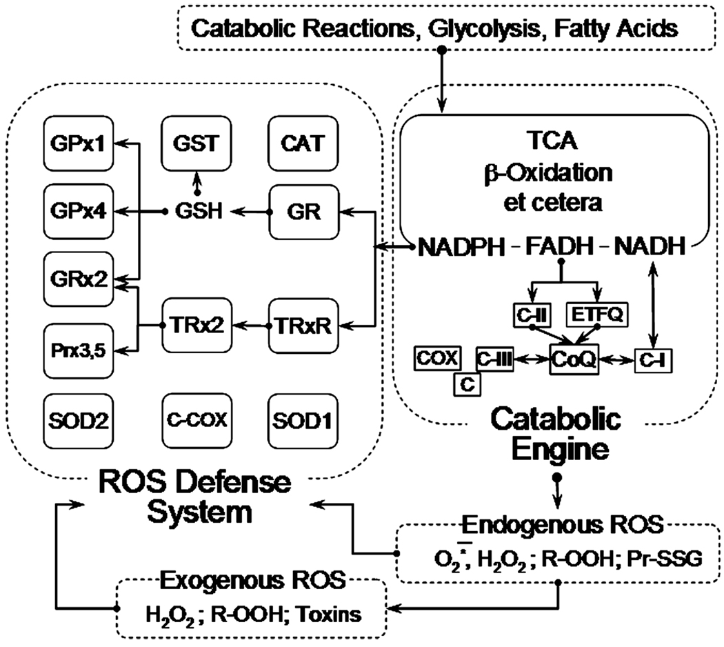
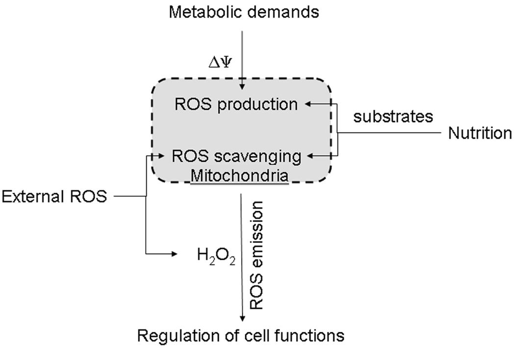
Oxidative stress is considered a major contributor to the etiology of both "normal" senescence and severe pathologies with serious public health implications. Several cellular sources, including mitochondria, are known to produce significant amounts of reactive oxygen species (ROS) that may contribute to intracellular oxidative stress. Mitochondria possess at least 10 known sites that are capable of generating ROS, but they also feature a sophisticated multilayered ROS defense system that is much less studied. This review summarizes the current knowledge about major components involved in mitochondrial ROS metabolism and factors that regulate ROS generation and removal at the level of mitochondria. An integrative systemic approach is applied to analysis of mitochondrial ROS metabolism, which is "dissected" into ROS generation, ROS emission, and ROS scavenging. The in vitro ROS-producing capacity of several mitochondrial sites is compared in the metabolic context and the role of mitochondria in ROS-dependent intracellular signaling is discussed.


Conflict of interest statement

Conflicts of Interest

The author declares no conflicts of interest.

Figures

Figure 1
Figure 1
Mitochondrial catabolic engine and reactive oxygen species (ROS) defense system. See text and Ref. for further detail. Abbreviations: GPx1, mitochondrial glutathione peroxidase; GPx4, mitochondrial phospholipid hydroperoxide glutathione peroxidase; Grx2, glutaredoxin-2; Prx3,5, peroxiredoxins 3 and 5; SOD2, mitochondrial manganese superoxide dismutase; GST, glutathione-S-transferase; GSH, reduced glutathione; TRx2, thioredoxin-2; CCOX, cytochrome c plus cytochrome c oxidase; Cat, catalase; GR, gluthatione reductase; TRxR, mitochondrial thioredoxin-2 reductase; SOD1, CuZn superoxide dismutase; TCA, tricarboxylic acid cycle; C-II, succinate dehydrogenase; ETFQ, electron transferring flavoprotein:CoQ reductase; CoQ, coenzyme Q; CI, Complex I of the respiratory chain; C-III, Complex III of the respiratory chain; C, cytochrome c; COX, cytochrome c oxidase.
Figure 2
Figure 2
Reported mitochondrial sites capable of ROS generation. Known ROS-generating enzymes are shown in a context of their location within mitochondria. See text and Ref. for further detail. Abbreviations: OM, outer mitochondrial membrane; IM, inner mitochondrial membrane; MAO, mono amine oxidases A and B; b5, cytochrome b5 reductase; DHOH, dihydroorotate dehydrogenase; αGDH, α-glycerophosphate dehydrogenase; C-I, Complex I of the respiratory chain; CoQ, coenzyme Q; C-III, Complex III of the respiratory chain; C, cytochrome c; COX, cytochrome c oxidase; SDH, succinate dehydrogenase; ACO, aconitase; KGDHC, α-ketoglutarate dehydrogenase complex; PDHC, pyruvate dehydrogenase complex; e, electrons. Arrows indicate the direction of electron flux between the enzymes and CoQ.
Figure 3
Figure 3
Features of succinate-supported ROS emission in rat brain mitochondria. (A) Concentration dependence of succinate-supported ROS emission in rat brain mitochondria incubated under nonphosphorylating conditions. Shaded boxes indicate the range of tissue succinate concentrations typically observed under normoxic conditions (normoxia) and after 20–30 min of hypoxia ischemia. See text for further detail. (B) Malate inhibits succinate-supported ROS emission in rat brain mitochondria incubated under nonphosphorylating conditions. Experimental conditions: incubation buffer was composed of 125 mM KCl, 2 mM KH2PO4, 1 mM MgCl2, 0.2 mg/ml of fatty acids-free BSA, 20 mM HEPES (pH 7.2), 0.2 mM EGTA, and (A) pictured concentrations of succinate or (B) 5 mM succinate. Mitochondria were added at 0.25 mg/ml; t = 37°. The emission of H2O2 was estimated from the changes in fluorescence using an H2O2 detection mixture of 4 U/ml of horseradish peroxidase, 40 U/ml SOD, and 10 µM Amplex Red (Molecular Probes, Oregon, USA).
Figure 4
Figure 4
Effect of the oxygen tension on the rates of phosphorylating respiration and H2O2 emission in rat liver mitochondria. The graph was rederived from the data reported in Ref. . See text for the detail.
Figure 5
Figure 5
Mitochondria sense changes in metabolism and ROS pressure and respond with an integrated signal in the form of H2O2. The magnitude of the mitochondrial membrane potential (ΔΨ) changes following fluctuations in the demands, such as ATP synthesis, which are imposed by cellular metabolism on the mitochondrial catabolic engine. Changes in cell nutrition are reflected by changes in the pattern of available oxidative substrates, monoamine oxidase substrates, and other factors. This results in changes in ROS generation in various mitochondrial sites. Internally generated ROS are combined with extramitochondrial ROS and get filtered through the mitochondrial ROS-scavenging system. The resulting level of the H2O2 emission serves as an integrated feedback signal from mitochondria to other intracellular H2O2-sensing systems.

References

    1. Lin MT, Beal MF. Mitochondrial dysfunction and oxidative stress in neurodegenerative diseases. Nature. 2006;443:787–795. - PubMed
    1. Orrenius S. Reactive oxygen species in mitochondria-mediated cell death. Drug. Metab. Rev. 2007;39:443–455. - PubMed
    1. Droge W, Schipper HM. Oxidative stress and aberrant signaling in aging and cognitive decline. Aging Cell. 2007;6:361–370. - PMC - PubMed
    1. Newsholme P. Diabetes associated cell stress and dysfunction: role of mitochondrial and non-mitochondrial ROS production and activity. J. Physiol. 2007;583:9–24. - PMC - PubMed
    1. Kim GJ, Chandrasekaran K, Morgan WF. Mitochondrial dysfunction, persistently elevated levels of reactive oxygen species and radiation-induced genomic instability: a review. Mutagenesis. 2006;21:361–367. - PubMed

Publication types

Substances