

Review
doi: 10.1196/annals.1427.020.
Structural diversity of mitochondria: functional implications
Affiliations
- PMID: 19076440
- PMCID: PMC2605638
- DOI: 10.1196/annals.1427.020
Item in Clipboard
Review
Structural diversity of mitochondria: functional implications
Ann N Y Acad Sci.
2008 Dec.
Abstract
Mitochondria display considerable structural diversity particularly in terms of the folding of the energy-transducing inner membrane. The hypothesis is forwarded that the topology of the mitochondrial inner membrane is not random but rather is a regulated cell parameter. Its effects on internal diffusion of metabolites and soluble proteins can influence such mitochondrial processes as ATP production and protein release during apoptosis. Progress toward understanding the factors that control mitochondrial inner-membrane curvature and dynamics (fusion and fission) is summarized, with a focus on remodeling events associated with apoptosis and oxidative stress.
Figures
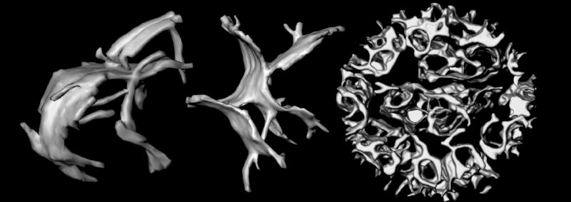
Topology of mitochondrial inner membranes. Left and middle: cristae of isolated, intact, frozen-hydrated rat-liver mitochondria. The large crista compartments appear to be formed by fusion of tubular membranes. These cristae are approximately 600 nm in length. Right: Inner membrane of a mouse liver mitochondrion pre-treated with the pro-apoptotic protein t-Bid. Rather than forming discrete compartments, the intracristal space is essentially one continuous compartment. The diameter of this mitochondrion is 860 nm
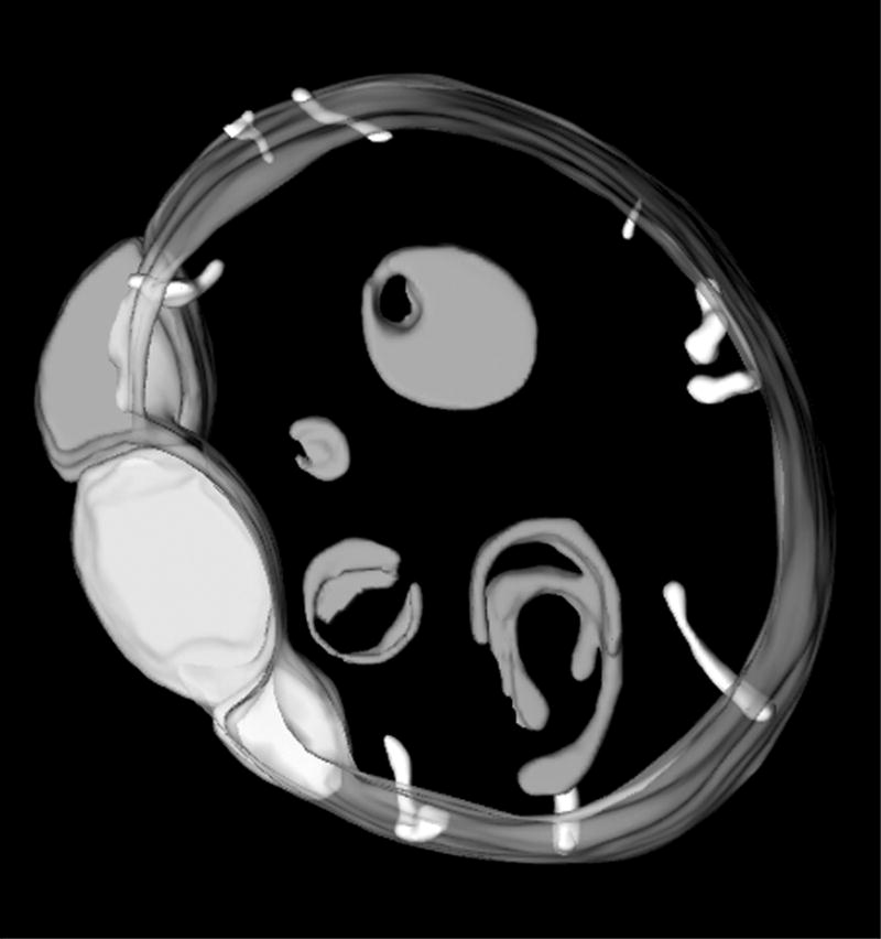
Internal mitochondrial membranes of an OPA1 deficient mutant of C. elegans. These mitochondria are characterized by numerous vesicular compartments, characteristic of a membrane fusion defect. The group of close-packed inner membrane vesicles at left is similar to features seen in apoptotic mitochondria. The diameter of this mitochondrion is 1.6 μm.
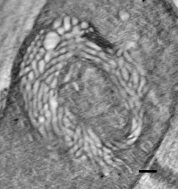
Slice from a tomogram of a “swirl” mitochondrion from Drosophila flight muscle., Tomographic analysis indicates that the swirl region is comprised of dilated, interconnected tubular cristae. (Specimen provided by Drs. S. Benzer and D. Walker.) The diameter of the mitochondrion is 900 nm.
References
-
- Munn EA. The Structure of Mitochondria. Academic Press; London: 1974.
-
- Fawcett DW. An Atlas of Fine Structure. W.B. Saunders Co.; Philadelphia: 1966.
-
- Tedeschi H, Harris DL. The osmotic behavior and permeability to non-electrolytes of mitochondria. Arch Biochem Biophys. 1955;58:52–67. - PubMed
Publication types
MeSH terms
Grants and funding
LinkOut - more resources
Full Text Sources
