. 2009 Jan;156(2):297-306.
doi: 10.1111/j.1476-5381.2008.00019.x. Epub 2008 Dec 9.

In vitro induction of T cells that are resistant to A2 adenosine receptor-mediated immunosuppression

Affiliations

In vitro induction of T cells that are resistant to A2 adenosine receptor-mediated immunosuppression

Akio Ohta et al. Br J Pharmacol. 2009 Jan.

Abstract

Background and purpose: The increased levels of extracellular adenosine in inflamed tissues down-regulate activated immune cells via the A(2A) adenosine receptor. This A(2A) adenosine receptor-mediated immunosuppression is a disqualifying obstacle in cancer immunotherapy as it protects cancerous tissues from adoptively transferred anti-tumour T cells. The aim of this study was to test whether the negative selection of T cells will produce T cells that are resistant to inhibition by extracellular adenosine.

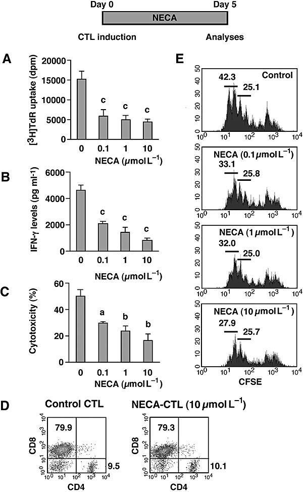
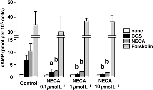
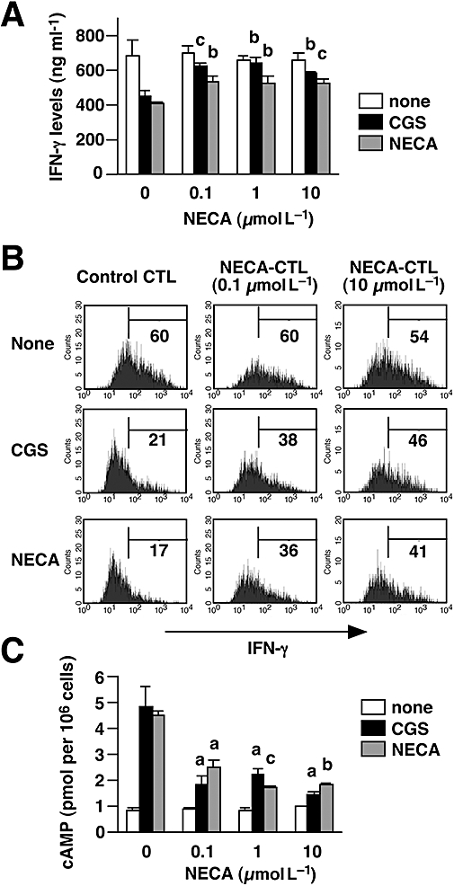
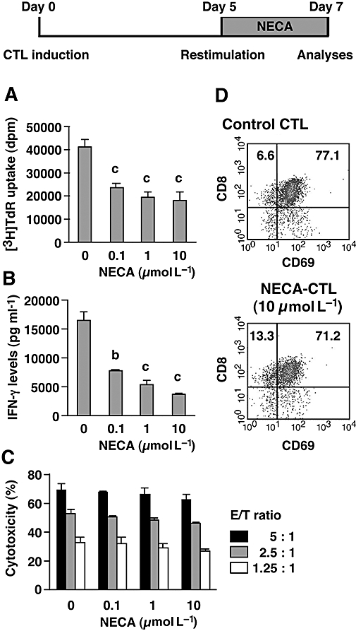
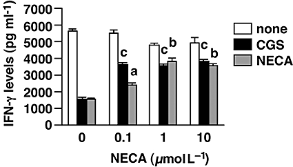
Experimental approach: Cytotoxic T lymphocytes (CTL) were developed by mixed lymphocyte culture in the presence or absence of the adenosine receptor agonist 5'-N-ethylcarboxamidoadenosine (NECA). The sensitivity of CTL to adenosine analogues was characterized by cAMP induction, interferon-gamma production and cytotoxicity.

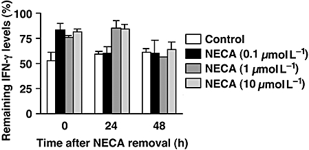
Key results: CTL that could proliferate even in the presence of NECA were less susceptible to inhibition by A(2A) adenosine receptor agonists, as shown by a much smaller accumulation of cAMP and less inhibition of interferon-gamma production compared with control CTL. The successful protocol to produce CTL that are both resistant to adenosine-mediated immunosuppression and maintain strong cytotoxicity and interferon-gamma secretion required NECA to be added only during the expansion stage after the establishment of CTL. In contrast, the priming of resting T cells in the presence of NECA resulted in T cells with impaired effector functions.

Conclusions and implications: Adenosine-resistant effector T cells were successfully obtained by exposure of activated T cells to NECA. These in vitro studies form the basis for future attempts to produce anti-tumour T cells that are more effective in adoptive immunotherapy.


Figures

Figure 1
Figure 1
NECA suppressed the mixed lymphocyte reaction, but there was significant development of CTL. CTL were induced by mixed lymphocyte culture using spleen cells from C57BL/6 (H-2b) and DBA/2 (H-2d) mice. The cells were cultured for 5 days in the presence or absence of NECA (0.1–10 µmol L−1). Resulting cells were examined for their proliferative activity (A), IFN-γ levels in the culture supernatant (B) and cytotoxicity against P815 mastocytoma (C). Effector–target ratio for cytotoxicity assay was 5:1. D. Proportion of CD4 and CD8 cells after 5 days. Large (activated) cells were gated for the analysis. Data from CTL induced with 10 µmol L−1 NECA shown are representative results; there was no marked difference in CTL induced with other concentrations of NECA. E. CD8+ cell expansion during mixed lymphocyte culture. C57BL/6 cells were stimulated after being labelled with CFSE and its fluorescence in CD8+ cells was analysed after 3 days of culture. Large (activated) CD8-expressing cells were gated for the analysis. Numbers in the panels indicate percentage of population. Data shown here represent average ± s.d. of triplicate samples. The statistical significance was calculated by Student's t-test: a P < 0.05; b P < 0.01; c P < 0.001. CFSE, carboxyfluorescein succinimidyl ester; CTL, cytotoxic T lymphocytes; IFN-γ, interferon-γ; NECA, 5′-N-ethylcarboxamidoadenosine; NECA-CTL, CTL developed in the presence of NECA.
Figure 2
Figure 2
CTL developed with NECA show impaired response to A2A/A2B adenosine receptor agonists. CTL were induced as described in Figure 1. cAMP production from the CTL was determined after incubation with A2A/A2B adenosine receptor agonists (CGS and NECA) and adenylate cyclase activator (forskolin). The concentration of cAMP inducers was 10 µmol L−1. Data shown here represent average ± s.d. of triplicate samples. The statistical significance was calculated by Student's t-test: a P < 0.05; b P < 0.01 versus control CTL. CGS, CGS21680; CTL, cytotoxic T lymphocytes; NECA, 5′-N-ethylcarboxamidoadenosine.
Figure 4
Figure 4
CTL treated with NECA for only the last 2 days were not only resistant to immunosuppression by A2A/A2B adenosine receptor agonists but also high IFN-γ producers comparable with control CTL. CTL were prepared as described for Figure 3. A and B. On day 7, the cells were re-stimulated by immobilized anti-CD3 and anti-CD28 mAbs for 24 h with CGS or NECA (10 µmol L−1). A. IFN-γ levels in the culture supernatant were quantified by ELISA. B. Intracellular IFN-γ expression in stimulated CTL was evaluated after further incubation with brefeldin A for 2 h. Numbers in each panel show the percentage of IFN-γ producers. The data shown here represent two separate experiments. C. NECA-CTL treated with NECA for only the last 2 days also showed an impaired cAMP response to A2A/A2B adenosine receptor agonists. Data shown here represent average ± s.d. of triplicate samples. The statistical significance was calculated by Student's t-test: a P < 0.05; b P < 0.01; c P < 0.001 versus control CTL. CTL, cytotoxic T lymphocytes; IFN-γ, interferon-γ; NECA, 5′-N-ethylcarboxamidoadenosine; NECA-CTL, CTL developed in the presence of NECA.
Figure 3
Figure 3
Cytotoxicity of NECA-CTL was maintained well when NECA was added only during the secondary mixed lymphocyte culture. Primary CTL were induced by mixed lymphocyte culture for 5 days. After extensive washing, the cells were re-stimulated with spleen cells from DBA/2 mice for 2 more days. NECA (0.1–1 µmol L−1) was withheld during the primary mixed lymphocyte culture and was added for 2 days after re-stimulation. The resulting cells were examined for their proliferative activity (A), IFN-γ levels in the culture supernatant (B), cytotoxicity against P815 mastocytoma (C) and flowcytometric analysis (D). Effector–target ratio for cytotoxicity assay was 5:1, 2.5:1 and 1.25:1. Data shown here represent average ± s.d. of triplicate samples. The statistical significance was calculated by Student's t-test: b P < 0.01; c P < 0.001. CTL, cytotoxic T lymphocytes; IFN-γ, interferon-γ; NECA, 5′-N-ethylcarboxamidoadenosine; NECA-CTL, CTL developed in the presence of NECA.
Figure 5
Figure 5
NECA-CTL maintained their resistance to A2A adenosine receptor stimulation at least for 24 h. NECA was removed by extensive washing 2 days after re-stimulation. Cells were re-cultured with IL-2 for 24–48 h, and then IFN-γ production was induced by immobilized anti-CD3 and anti-CD28 mAbs. The inhibitory effect of CGS (10 µmol L−1) is expressed as a percentage of IFN-γ levels remaining compared with control. Data shown represent average ± s.d. of triplicate samples. CTL, cytotoxic T lymphocytes; IFN-γ, interferon-γ; NECA, 5′-N-ethylcarboxamidoadenosine; NECA-CTL, CTL developed in the presence of NECA.
Figure 6
Figure 6
NECA-CTL could produce high levels of IFN-γ upon recognition of tumour cells even in the presence of A2A/A2B adenosine receptor agonists. CTL were induced as described for Figure 3. In order to activate antigen-specific anti-tumour responses, the same number of CTL was evaluated after co-culture with mitomycin C-treated P815 cells. The susceptibility of the CTL to A2A/A2B adenosine receptor activation was examined by stimulating them in the presence of CGS or NECA (10 µmol L−1). IFN-γ levels in the supernatant were determined after 2 days. Data shown here represent average ± s.d. of triplicate samples. The statistical significance was calculated by Student's t-test: a P < 0.05; b P < 0.01; c P < 0.001 versus control CTL. CGS, CGS21680; CTL, cytotoxic T lymphocytes; IFN-γ, interferon-γ; NECA, 5′-N-ethylcarboxamidoadenosine; NECA-CTL, CTL developed in the presence of NECA.

Similar articles

Cited by

References

    1. Annacker O, Pimenta-Araujo R, Burlen-Defranoux O, Barbosa TC, Cumano A, Bandeira A. CD25+ CD4+ T cells regulate the expansion of peripheral CD4 T cells through the production of IL-10. J Immunol. 2001;166:3008–3018. - PubMed
    1. Antony PA, Piccirillo CA, Akpinarli A, Finkelstein SE, Speiss PJ, Surman DR, et al. CD8+ T cell immunity against a tumor/self-antigen is augmented by CD4+ T helper cells and hindered by naturally occurring T regulatory cells. J Immunol. 2005;174:2591–2601. - PMC - PubMed
    1. Apasov SG, Chen JF, Smith PT, Schwarzschild MA, Fink JS, Sitkovsky MV. Study of A(2A) adenosine receptor gene deficient mice reveals that adenosine analogue CGS 21680 possesses no A(2A) receptor-unrelated lymphotoxicity. Br J Pharmacol. 2000;131:43–50. - PMC - PubMed
    1. Asseman C, Mauze S, Leach MW, Coffman RL, Powrie F. An essential role for interleukin 10 in the function of regulatory T cells that inhibit intestinal inflammation. J Exp Med. 1999;190:995–1004. - PMC - PubMed
    1. von Boehmer H. Mechanisms of suppression by suppressor T cells. Nat Immunol. 2005;6:338–344. - PubMed

Publication types

MeSH terms