. 2009 Mar;100(3):449-56.
doi: 10.1111/j.1349-7006.2008.01051.x. Epub 2008 Dec 14.

Critical role for constitutive type I interferon signaling in the prevention of cellular transformation

Affiliations

Critical role for constitutive type I interferon signaling in the prevention of cellular transformation

Hui-Min Chen et al. Cancer Sci. 2009 Mar.

Abstract

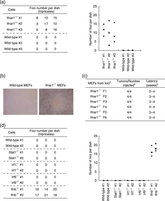
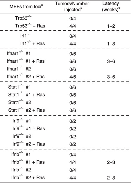
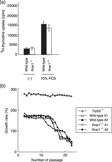
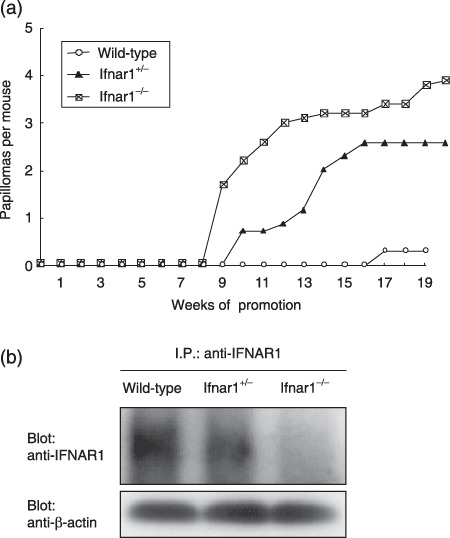
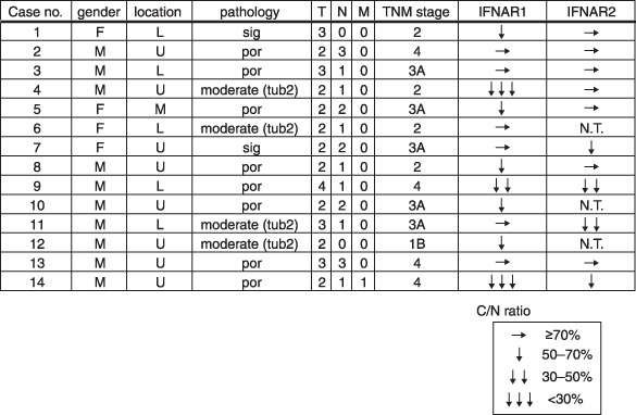
Interferons-alpha/beta, which are produced upon viral infection, are key soluble factors for the establishment of an antiviral state, but are also produced at low levels in the absence of infection. Herein, we demonstrate that a weak signal by these constitutively produced IFN-alpha/beta show a preventive role in cellular transformation. Ifnar1-deficient (Ifnar1(-/-)) MEF, which are devoid of IFN-alpha/beta signal, undergo a spontaneous transformation during long-term cell culture. Similar to Irf1(-/-) MEF, primary Ifnar1(-/-) MEF become tumorigenic in nude mice by the expression of activated c-Ha-Ras oncoprotein. However, Ifnar1(-/-) MEF do not show any abnormal growth properties. A similar observation is made in Ifnb(-/-) MEF that fail to produce constitutive IFN-alpha/beta, whereas such a transforming property is not found in MEF that lack any of the IFN receptor downstream molecules including Stat1, IRF9 and IRF1. Furthermore, Ifnar1(-/-) mice develop chemically-induced skin papilloma more severely than wild-type mice. In addition, the expression levels of IFNAR1 mRNA are significantly decreased in human gastric cancer tissues. These results suggest a cell-intrinsic role of the weak signal by constitutively produced IFN-alpha/beta to prevent cells from transformation, which may be mediated by a hitherto-unknown pathway(s) downstream of the IFN-alpha/beta receptor.


Figures

Figure 1
Figure 1
Spontaneous transformation of Ifnar1−/– MEF. (a) Primary wild‐type or Ifnar1−/– MEF (3 × 106 cells) were plated on 10‐cm culture dishes, and after 4‐ to 6‐week cell culture, numbers of foci formed in the dish were counted. Assay was done with three different MEF in three independent dishes. Average of the numbers of foci observed in the three dishes are shown as a horizontal line in the graph (right). (b) Appearance of a cell colony observed after 50‐day culture. (c) Six cell clones (F1–F6) were isolated from separate foci that formed after long‐term cell culture, were inoculated in nude mice for tumorigenicity. aMEF genotypes; bnumber of tumors arising per number of sites injected; cperiod until visible tumor formation. (d) Primary wild‐type or other mutant MEF lacking signaling molecules downstream of the IFNAR were plated on 10‐cm culture dishes, and were examined for the formation of colonies during the long‐term culture, as described in (a). IFNAR, IFN‐α/β receptor; MEF, mouse embryonic fibroblasts.
Figure 2
Figure 2
Oncogenic transformation of primary MEF deficient in IFNAR1, its downstream molecules, or IFN‐β, by the expression of c‐Ha‐Ras oncoprotein. In vivo tumorigenesis of mutant MEF lacking molecules involved in IFN‐α/β‐mediated signaling. Primary MEF, freshly prepared from Ifnar1−/– or ifnb−/– embryos, were infected with pGDV12ras retrovirus, which directs the expression of an activated form of c‐Ha‐Ras oncoprotein carrying an oncogenic G12V mutation.( 21 ) These cells were subjected to tumorigenesis assay in nude mice as described in Fig. 1 (c). IFN‐β, β‐interferon; IFNAR, IFN‐α/β receptor; MEF, mouse embryonic fibroblasts.
Figure 3
Figure 3
Normal growth properties of Ifnar1−/– MEF. (a) (3H)thymidine incorporation assay using primary wild‐type and Ifnar1−/– MEF. Indicated cells (1.0 × 105) of the indicated cells were plated and grown in media containing 10% FCS for 32 h followed by a (3H)thymidine pulse, and 16 h later, the (3H)thymidine uptake levels were measured by scintillation counting. Values of the average and standard deviation of three measures are shown. (b) Replicative senescence in Ifnar1−/– MEF. Wild‐type, Ifnar1−/– and Trp53−/– MEF were routinely cultured over a period of 23 passages. MEF from two different embryo preparations were used for each genotype (#1 and #2). Cells were counted at subculture to determine the population doublings. Wild‐type and Ifnar1−/– MEF but not Trp53−/– MEF reached replicative senescence after 12–14 passages. cpm, count per minute; FCS, fetal calf serum; MEF, mouse embryonic fibroblasts.
Figure 4
Figure 4
Irreversible transforming property of Ifnar1−/– MEFs. (a) Methylcellulose gel assay. Two randomly selected clones of either spontaneously transformed Ifnar1−/– MEFs (F1 and F2) or Ifnar1−/– MEFs transformed by activated Ras expression (#1 and #2) were infected with a retrovirus that directs the expression of mouse IFNAR1 (solid bars) and the control retrovirus (open bars). In this assay, a total of 5000 viable cells were counted under a microscope, and the number of colonies formed among them was scored. Values are indicated as percentage of total cells examined. (b) DNA binding activity of ISGF3 in transformed Ifnar1−/– MEF reconstituted by the expression of IFNAR1. The transformed Ifnar1−/– MEFs (F1 and #1) retrovirally expressing IFNAR1 or mock are stimulated with IFN‐β (500 U/mL), and subjected to EMSA by using ISRE probe. EMSA, electromobility shift assays; IFN‐β, β‐interferon; IFNAR, IFN‐α/β receptor; ISRE, interferon‐stimulated response element; MEF, mouse embryonic fibroblasts.
Figure 5
Figure 5
Development of skin papillomas in Ifnar1−/– mice. (a) Groups of wild‐type, Ifnar1+/– and Ifnar1−/– mice (10 mice per group) were initiated by a single treatment with DMBA and promoted twice a week with TPA for 15 weeks. The average number of papillomas (over 2 mm in diameter) per mouse was plotted every week after the start of carcinogen treatment. (b) Constitutive expression levels of IFNAR1 protein in the skin tissues of wild‐type, Ifnar1+/– and Ifnar1−/– mice. Immunoprecipitates with anti‐IFNAR1 were subjected to western blotting analysis with anti‐IFNAR1. The loading amount of each lane was also confirmed by Western blotting with an aliquot of whole cell lysates that were used for immunoprecipitation. DMBA, 7,12‐dimethylbenz(a)anthracene; TPA, 12‐O‐tetradecanoylphorbol 13‐acetate; IFNAR, α/β‐interferon receptor; I.P., Immunoprecipitation.
Figure 6
Figure 6
Decreased expression levels of the type I IFN receptor subunits in human gastric cancer tissues. Total RNA were extracted from cancerous and non‐cancerous tissues of 14 patients with various types of gastric cancers, and were analyzed by quantitative RT‐PCR with specific primers. C/N ratios were calculated by comparing the expression level of cancerous tissues with that of non‐cancerous tissue. L, lower region of the stomach; M, middle region of the stomach; N.T., not tested; por, poor differentiated carcinoma; RT‐PCR, reverse transcription polymerase chain reaction; sig, signet ring cell carcinoma; tub2, tubular adenocarcinoma (moderately differentiated type); U, upper region of the stomach.

References

    1. Novick D, Cohen B, Rubinstein M. The human interferon alpha/beta receptor: characterization and molecular cloning. Cell 1994; 77: 391–400. - PubMed
    1. Stark GR, Kerr IM, Williams BR, Silverman RH, Schreiber RD. How cells respond to interferons. Annu Rev Biochem 1998; 67: 227–64. - PubMed
    1. Uze G, Lutfalla G, Gresser I. Genetic transfer of a functional human interferon alpha receptor into mouse cells: cloning and expression of its cDNA. Cell 1990; 60: 225–34. - PubMed
    1. Decker T, Lew DJ, Darnell JE Jr. Two distinct α‐interferon‐dependent signal transduction pathways may contribute to activation of transcription of the guanylate‐binding protein gene. Mol Cell Biol 1991; 11: 5147–53. - PMC - PubMed
    1. Bluyssen AR, Durbin JE, Levy DE. ISGF3 gamma p48, a specificity switch for interferon activated transcription factors. Cytokine Growth Factor Rev 1996; 7: 11–7. - PubMed

Publication types

MeSH terms