

Giant cell formation in sarcoidosis: cell fusion or proliferation with non-division?
- PMID: 19077083
- PMCID: PMC2669524
- DOI: 10.1111/j.1365-2249.2008.03841.x
Giant cell formation in sarcoidosis: cell fusion or proliferation with non-division?
Abstract
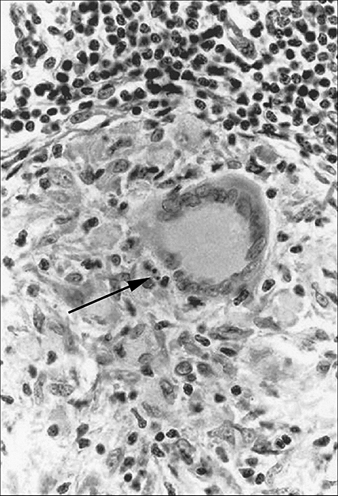
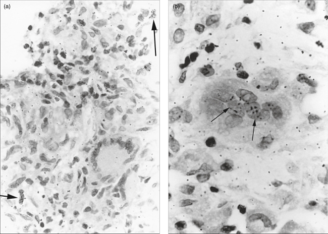
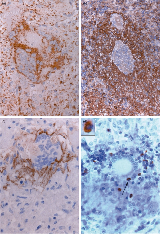
Granulomas are inflammatory reactions featuring macrophages, epithelioid, T and multi-nucleated giant cells (MGC). Giant cells are present in a number of granulomatous reactions, but little is known about their formation and function, especially in man. We studied MGC in the granulomatous disorder sarcoidosis. In situ labelling of lymph nodes by means of [(3)H]-thymidine showed that proliferation and non-division of epithelioid cells leading towards giant cells was not observed in these granulomas. However, [(3)H]-uridine incorporation showed MGC with labelled as well as unlabelled nuclei in the same cell, pointing to a process of fusion of epithelioid cells to form giant cells. Apoptotic bodies were incidentally found in granulomas. A novel finding was that such bodies were statistically more often found in the close vicinity of MGC, but not within these cells. These apoptotic cells appeared to be CD4(+) lymphocytes or histiocytes. CD44 and CCR-5 involved in the process of fusion were expressed in MGC. In conclusion, MGC in sarcoidosis derive by cell fusion rather than by proliferation and non-division, and seem to play an active role in the induction of apoptosis in granulomas.
Figures
References
-
- Chambers TJ. Multinucleate giant cells. J Pathol. 1978;126:125–48. - PubMed
-
- Thoenes W, Sonntag W, Heine WD, Langer KH. Cell fusion as a mechanism for the formation of giant cells (Langhans’ type). Autoradiographic findings in autoimmune tubulo-interstitial nephritis of the rat. Virchows Arch B. 1982;41:45–50. - PubMed
-
- Ryan GB, Spector WG. Macrophage turnover in inflamed connective tissue. Proc R Soc Lond B Biol Sci. 1970;175:269–92. - PubMed
-
- Mariano M, Spector WG. The formation and properties of macrophage polykaryons (inflammatory giant cells) J Pathol. 1974;113:1–19. - PubMed
MeSH terms
Substances
LinkOut - more resources
Full Text Sources
Medical
Research Materials
Miscellaneous
