. 2008 Dec 11:6:79.
doi: 10.1186/1479-5876-6-79.

Aurora kinase inhibitors synergize with paclitaxel to induce apoptosis in ovarian cancer cells

Affiliations

Aurora kinase inhibitors synergize with paclitaxel to induce apoptosis in ovarian cancer cells

Christopher D Scharer et al. J Transl Med. .

Abstract

Background: A large percentage of patients with recurrent ovarian cancer develop resistance to the taxane class of chemotherapeutics. While mechanisms of resistance are being discovered, novel treatment options and a better understanding of disease resistance are sorely needed. The mitotic kinase Aurora-A directly regulates cellular processes targeted by the taxanes and is overexpressed in several malignancies, including ovarian cancer. Recent data has shown that overexpression of Aurora-A can confer resistance to the taxane paclitaxel.

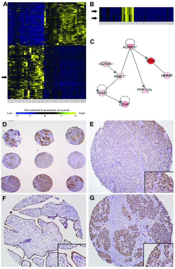
Methods: We used expression profiling of ovarian tumor samples to determine the most significantly overexpressed genes. In this study we sought to determine if chemical inhibition of the Aurora kinase family using VE-465 could synergize with paclitaxel to induce apoptosis in paclitaxel-resistant and sensitive ovarian cancer cells.

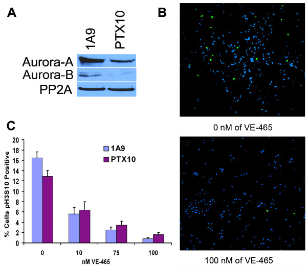
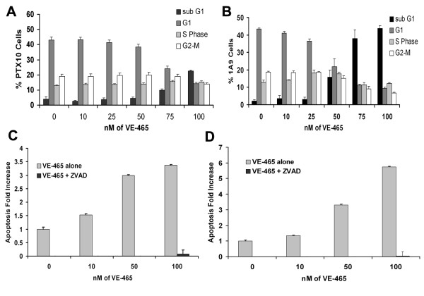
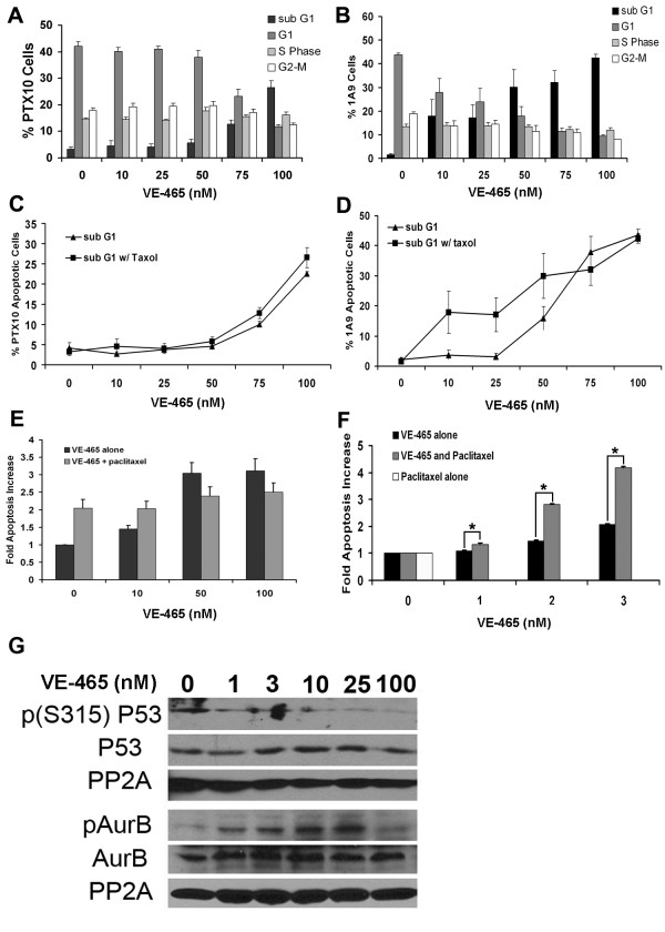
Results: Aurora-A kinase and TPX2, an activator of Aurora-A, are two of the most significantly overexpressed genes in ovarian carcinomas. We show that inhibition of the Aurora kinases prevents phosphorylation of a mitotic marker and demonstrate a dose-dependent increase of apoptosis in treated ovarian cancer cells. We demonstrate at low doses that are specific to Aurora-A, VE-465 synergizes with paclitaxel to induce 4.5-fold greater apoptosis than paclitaxel alone in 1A9 cells. Higher doses are needed to induce apoptosis in paclitaxel-resistant PTX10 cells.

Conclusion: Our results show that VE-465 is a potent killer of taxane resistant ovarian cancer cells and can synergize with paclitaxel at low doses. These data suggest patients whose tumors exhibit high Aurora-A expression may benefit from a combination therapy of taxanes and Aurora-A inhibition.


Figures

Figure 1
Figure 1
Aurora-A is overexpressed in carcinomas. Heat map image of Z-score normalized microarray expression data from Affymetrix U95A gene chips. Genes with lower expression compared to normal tissue are shown in blue and yellow indicates genes that are overexpressed. (A) Heat map representing the entire data set. Arrow indicates Aurora-A. (B) Aurora-A is overexpressed 5 fold in carcinomas compared to adenomas. Both Aurora-A probes are shown. Ca – carcinoma, Ad – adenoma, CC – cancers pre-treated with chemotherapy. (C) Ingenuity Pathway Assist analysis of significantly overexpressed genes. Diagram represents an interaction network of the 8 genes and Aurora-A kinase. (D) Low power (2×) image of ovarian tissue microarray stained for Aurora A by immunohistochemistry. (E) Aurora-A staining of TMA core of ovarian carcinoma without adjuvant chemotherapy (20×). (F) Aurora-A staining of TMA core of benign ovarian tissue (20×). (G) Aurora-A staining of TMA core of ovarian carcinoma with adjuvant chemotherapy (20×).
Figure 2
Figure 2
VE-465 inhibits the Aurora kinases. (A) Immunoblot analysis of whole cell lysates from 1A9 and PTX10 cell lines probed for Aurora-A, Aurora-B and PP2A as a loading control. (B) Paclitaxel-resistant PTX10 and IA9 cells were treated for 48 hours with VE-465. Following treatment, mitotic cells were assessed by staining for Histone H3 phosphorylated on Ser10 (pH3S10), a marker of mitosis and an Aurora-B substrate (green). Nuclear chromatin was visualized with the To-Pro (blue) counter stain to indicate total number of cells. (C) Ten random fields were sampled for each concentration and percentage of pH3S10 positive cells calculated.
Figure 3
Figure 3
Inhibition of Aurora kinases results in cell death. Cells were treated for 96 hours with differing doses of VE-465. (A) PTX10 cells (B) 1A9 cells. Following treatment cells were harvested, fixed and stained with propidium iodide before analysis by Flow Cytometry. The sub G0/G1 population represents apoptotic cells. Each time point represents data from at least 3 independent experiments. Caspase 3/7 assays of PTX10 (C) and 1A9 (D) cells treated with increasing doses of VE-465 demonstrate dose-dependent increase in apoptosis. The caspase activity was blocked by the pan-caspase inhibitor Z-VAD.
Figure 4
Figure 4
VE-465 induces cell death in the presence of paclitaxel. Cells were treated for 96 hours with differing doses of VE-465 in the presence of 15 ng/mL paclitaxel. (A) PTX10 cells (B) 1A9 cells. Analysis was performed as described in Figure 3. The sub G0/G1 population represents apoptotic cells. Each time point represents data from at least 3 independent experiments. Paclitaxel and VE-465 did not synergize to cause apoptosis in PTX10 (C) or 1A9 (D) cells. Percent of apoptotic cells are plotted for cells treated for 96 hrs with VE-465 alone or VE-465 and 15 ng/mL paclitaxel. Triangles – cells treated with increasing concentrations of VE-465. Squares – cells treated with increasing concentrations of VE-465 in the presence of 15 ng/mL paclitaxel. (E) Caspase 3/7 assays of PTX10 cells treated with 10–100 nM of VE-465 alone or in combination with 15 ng/mL paclitaxel. Confirming flow cytometry data, combination treatment with paclitaxel and VE-465 did not synergistically increase apoptosis in the PTX10 cell line. (F) Caspase 3/7 assays of 1A9 cells treated with 1–3 nM of VE-465 alone, 15 ng/mL paclitaxel alone, or in combination with 15 ng/mL paclitaxel. A dose of 3 nM VE-465 alone induced 2-fold more apoptosis than 15 ng/mL paclitaxel, whereas combined 3 nM VE-465 and 15 ng/mL paclitaxel synergistically induced 4.5-fold more apoptosis than 15 ng/mL paclitaxel alone. (* = p-value less than 0.0025 by students T-test.) (G) Immunoblot of 1A9 cells treated with increasing concentrations of VE-465 for 96 hours. The kinase activity of Aurora-A and Aurora-B is suppressed in a dose-dependent manner consistent with the known Ki values of VE-465. Phosphorylation of the Aurora-A target p53 (S315) is inhibited at doses of 1 nM and higher whereas auto-phosphorylation of Aurora-B (T232) is only inhibited at doses exceeding 25 nM.

Similar articles

Cited by

References

    1. Weaver BA, Cleveland DW. Decoding the links between mitosis, cancer, and chemotherapy: The mitotic checkpoint, adaptation, and cell death. Cancer Cell. 2005;8:7–12. doi: 10.1016/j.ccr.2005.06.011. - DOI - PubMed
    1. Marumoto T, Zhang D, Saya H. Aurora-A – a guardian of poles. Nat Rev Cancer. 2005;5:42–50. doi: 10.1038/nrc1526. - DOI - PubMed
    1. Glover DM, Leibowitz MH, McLean DA, Parry H. Mutations in aurora prevent centrosome separation leading to the formation of monopolar spindles. Cell. 1995;81:95–105. doi: 10.1016/0092-8674(95)90374-7. - DOI - PubMed
    1. Keen N, Taylor S. Aurora-kinase inhibitors as anticancer agents. Nat Rev Cancer. 2004;4:927–936. doi: 10.1038/nrc1502. - DOI - PubMed
    1. Adams RR, Maiato H, Earnshaw WC, Carmena M. Essential roles of Drosophila inner centromere protein (INCENP) and aurora B in histone H3 phosphorylation, metaphase chromosome alignment, kinetochore disjunction, and chromosome segregation. J Cell Biol. 2001;153:865–880. doi: 10.1083/jcb.153.4.865. - DOI - PMC - PubMed

Publication types

MeSH terms