. 2008 Dec 11:5:146.
doi: 10.1186/1743-422X-5-146.

Occult hepatitis B infection: an evolutionary scenario

Affiliations

Occult hepatitis B infection: an evolutionary scenario

Formijn J van Hemert et al. Virol J. .

Abstract

Background: Occult or latent hepatitis B virus (HBV) infection is defined as infection with detectable HBV DNA and undetectable surface antigen (HBsAg) in patients' blood. The cause of an overt HBV infection becoming an occult one is unknown. To gain insight into the mechanism of the development of occult infection, we compared the full-length HBV genome from a blood donor carrying an occult infection (d4) with global genotype D genomes.

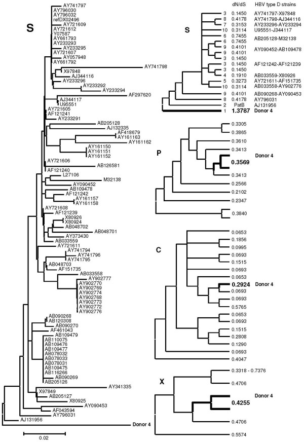
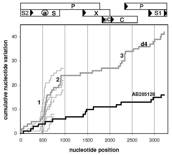
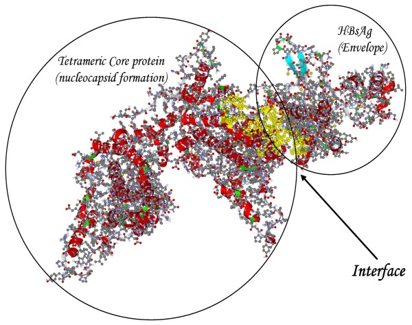
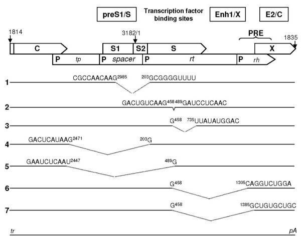
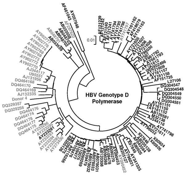
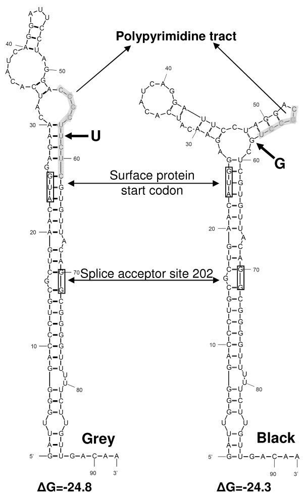
Results: The phylogenetic analysis of polymerase, core and X protein sequences did not distinguish d4 from other genotype D strains. Yet, d4 surface protein formed the evolutionary outgroup relative to all other genotype D strains. Its evolutionary branch was the only one where accumulation of substitutions suggests positive selection (dN/dS = 1.3787). Many of these substitutions accumulated specifically in regions encoding the core/surface protein interface, as revealed in a 3D-modeled protein complex. We identified a novel RNA splicing event (deleting nucleotides 2986-202) that abolishes surface protein gene expression without affecting polymerase, core and X-protein related functions. Genotype D strains differ in their ability to perform this 2986-202 splicing. Strains prone to 2986-202 splicing constitute a separate clade in a phylogenetic tree of genotype D HBVs. A single substitution (G173T) that is associated with clade membership alters the local RNA secondary structure and is proposed to affect splicing efficiency at the 202 acceptor site.

Conclusion: We propose an evolutionary scenario for occult HBV infection, in which 2986-202 splicing generates intracellular virus particles devoid of surface protein, which subsequently accumulates mutations due to relaxation of coding constraints. Such viruses are deficient of autonomous propagation and cannot leave the host cell until it is lysed.


Figures

Figure 1
Figure 1
HBV strain phylogeny. A bootstrap consensus tree based on JTT-estimates of amino acid replacements in surface proteins of HBV genotype D displays the surface protein of donor 4 carrying an occult infection in the outgroup position (left panel). The scale bar indicates 2% of evolutionary divergence. For phylogenetic analysis by maximum likelihood, the HBV type D strains were grouped according to their topological position, approximately and provided with labels as indicated next to the branches of the compressed topology tree (right panel, S). The corresponding values obtained for dN/dS are in between of the labels and strains columns; PatB means ''parameter at boundary''. Data on donor 4 are in bold-face. The three panels marked by P(olymerase), C(ore) and X were constructed in a similar fashion, but without mentioning GenBank IDs and clade/branch labels. In case of P and X, the donor 4 species was combined with its nearest neighbor in order to avoid deviation due to insufficient branch length.
Figure 2
Figure 2
Mutational scan along the HBV genome. Nucleotide substitutions uniquely present in EU155893 HBV DNA (d4, thick grey line, occult infection) and in control AB205128 HBV DNA (thick black line, overt infection) are compared with 89 HBV DNAs of genotype D and plotted cumulatively along the HBV genome. Steep slopes at the a-determinant (1), the oligonucleotide 895–909 (2) and the central part of C (3) indicate the relatively high divergence of these regions in d4 HBV. Thin grey lines represent characteristic mutations in the available HBV sequences from blood samples of the other donors with occult HBV infection. Numbering starts from the conventional EcoR1 site between S1 and S2. A map of HBV genome organization is provided on top of the figure.
Figure 3
Figure 3
Model of the core/surface protein interaction. A 3D-modeled complex of tetrameric core protein with HBsAg monomer shows the yellow-colored amino acid residues comprising the interface between the two proteins.
Figure 4
Figure 4
RNA splicing possibilities in the HBV genome. Splice patterns in HBV pregenomic RNA are predicted by NetGene2. Genome organization is presented as functional domains in pgRNA decorated with transcription factor binding sites (upper two panels) from terminal repeat (tr) to polyadenylation sequence (pA) (bottom line). PRE indicates the position of the Posttranscriptional Regulatory Element. Different modes of splicing are numbered and referred to in the text.
Figure 5
Figure 5
Detailed phylogeny of HBV genotype D strains. A phylogenetic bootstrapped consensus tree of HBV genotype D strains was derived from replacements in the amino acid sequences of the viral polymerase. Grey clade members scored positively with respect to the 202 acceptor site predicted by NetGene2, in contrast with members of the black collection. The scale bar indicates 1% of evolutionary divergence.
Figure 6
Figure 6
Analysis of splice acceptor site 202 in the HBV genome. A single U173G mutation affects the local RNA secondary structure. A consensus sequence of grey clade members (left panel) differs from the black collection (right panel) by an U-versus a G-nucleotide promoting exposure into a loop structure or burial into a stem structure, respectively, of a polypyrimidine tract (marked by shading) obligatory for efficient splicing at the 202 acceptor site indicated by an arrow. For the purpose of orientation, the AUG initiation codon for surface protein translation is also indicated. Values for ΔG are in kcal/mole.

Similar articles

Cited by

References

    1. Raimondo G, Pollicino T, Cacciola I, Squadrito G. Occult hepatitis B virus infection. J Hepatol. 2007;46:160–170. doi: 10.1016/j.jhep.2006.10.007. - DOI - PubMed
    1. Jeantet D, Chemin I, Mandrand B, Tran A, Zoulim F, Merle P, Trepo C, Kay A. Cloning and expression of surface antigens from occult chronic hepatitis B virus infections and their recognition by commercial detection assays. J Med Virol. 2004;73:508–515. doi: 10.1002/jmv.20119. - DOI - PubMed
    1. Garfein RS, Bower WA, Loney CM, Hutin YJ, Xia GL, Jawanda J, Groom AV, Nainan OV, Murphy JS, Bell BP. Factors associated with fulminant liver failure during an outbreak among injection drug users with acute hepatitis B. Hepatology. 2004;40:865–873. - PubMed
    1. Petzold DR, Tautz B, Wolf F, Drescher J. Infection chains and evolution rates of hepatitis B virus in cardiac transplant recipients infected nosocomially. J Med Virol. 1999;58:1–10. doi: 10.1002/(SICI)1096-9071(199905)58:1<1::AID-JMV1>3.0.CO;2-M. - DOI - PubMed
    1. Gerlich WH, Wagner FF, Chudy M, Harritshoj LH, Lattermann A, Wienzek S, Glebe D, Saniewski M, Schüttler CG, Wend UC, et al. HBsAg Non-Reactive HBV Infection in Blood Donors: Transmission and Pathogenicity. J Med Virol. 2007;79:S32–S36. doi: 10.1002/jmv.20963. - DOI

MeSH terms